Calcium »
PDB 1ukg-1ux6 »
1uoc »
Calcium in PDB 1uoc: X-Ray Structure of the Rnase Domain of the Yeast POP2 Protein
Protein crystallography data
The structure of X-Ray Structure of the Rnase Domain of the Yeast POP2 Protein, PDB code: 1uoc
was solved by
S.Thore,
F.Mauxion,
B.Seraphin,
D.Suck,
with X-Ray Crystallography technique. A brief refinement statistics is given in the table below:
| Resolution Low / High (Å) | 9.97 / 2.3 |
| Space group | P 21 21 21 |
| Cell size a, b, c (Å), α, β, γ (°) | 78.582, 79.440, 101.324, 90.00, 90.00, 90.00 |
| R / Rfree (%) | 23.9 / 26.5 |
Other elements in 1uoc:
The structure of X-Ray Structure of the Rnase Domain of the Yeast POP2 Protein also contains other interesting chemical elements:
| Xenon | (Xe) | 2 atoms |
Calcium Binding Sites:
The binding sites of Calcium atom in the X-Ray Structure of the Rnase Domain of the Yeast POP2 Protein
(pdb code 1uoc). This binding sites where shown within
5.0 Angstroms radius around Calcium atom.
In total 2 binding sites of Calcium where determined in the X-Ray Structure of the Rnase Domain of the Yeast POP2 Protein, PDB code: 1uoc:
Jump to Calcium binding site number: 1; 2;
In total 2 binding sites of Calcium where determined in the X-Ray Structure of the Rnase Domain of the Yeast POP2 Protein, PDB code: 1uoc:
Jump to Calcium binding site number: 1; 2;
Calcium binding site 1 out of 2 in 1uoc
Go back to
Calcium binding site 1 out
of 2 in the X-Ray Structure of the Rnase Domain of the Yeast POP2 Protein
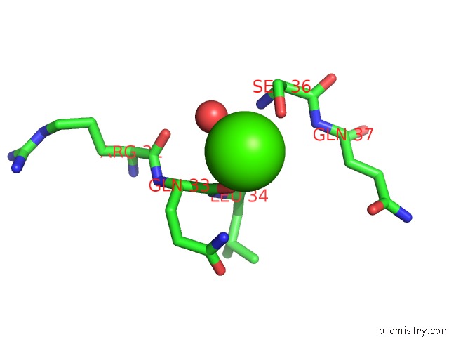
Mono view
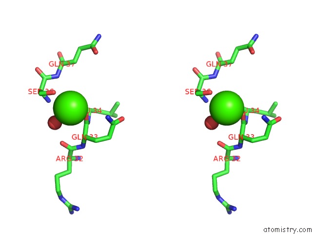
Stereo pair view
Mono view
Stereo pair view
A full contact list of Calcium with other atoms in the Ca binding
site number 1 of X-Ray Structure of the Rnase Domain of the Yeast POP2 Protein within 5.0Å range:
|
Calcium binding site 2 out of 2 in 1uoc
Go back to
Calcium binding site 2 out
of 2 in the X-Ray Structure of the Rnase Domain of the Yeast POP2 Protein
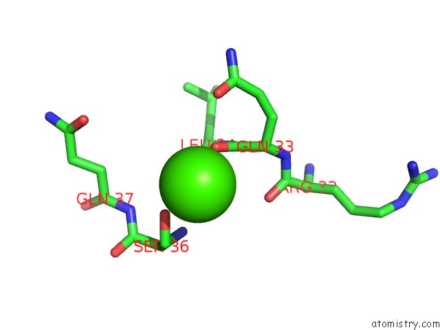
Mono view
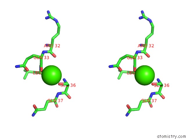
Stereo pair view
Mono view
Stereo pair view
A full contact list of Calcium with other atoms in the Ca binding
site number 2 of X-Ray Structure of the Rnase Domain of the Yeast POP2 Protein within 5.0Å range:
|
Reference:
S.Thore,
F.Mauxion,
B.Seraphin,
D.Suck.
X-Ray Structure and Activity of the Yeast POP2 Protein: A Nuclease Subunit of the Mrna Deadenylase Complex Embo Rep. V. 4 1150 2003.
ISSN: ISSN 1469-221X
PubMed: 14618157
DOI: 10.1038/SJ.EMBOR.7400020
Page generated: Tue Jul 8 02:37:15 2025
ISSN: ISSN 1469-221X
PubMed: 14618157
DOI: 10.1038/SJ.EMBOR.7400020
Last articles
Cl in 5RAQCl in 5RAP
Cl in 5RAO
Cl in 5RAN
Cl in 5RAJ
Cl in 5RAM
Cl in 5RAL
Cl in 5RAK
Cl in 5RAF
Cl in 5RAG