Calcium »
PDB 1ukg-1ux6 »
1us1 »
Calcium in PDB 1us1: Crystal Structure of Human Vascular Adhesion Protein-1
Enzymatic activity of Crystal Structure of Human Vascular Adhesion Protein-1
All present enzymatic activity of Crystal Structure of Human Vascular Adhesion Protein-1:
1.4.3.6;
1.4.3.6;
Protein crystallography data
The structure of Crystal Structure of Human Vascular Adhesion Protein-1, PDB code: 1us1
was solved by
T.T.Airenne,
Y.Nymalm,
H.Kidron,
A.Soderholm,
M.S.Johnson,
T.A.Salminen,
with X-Ray Crystallography technique. A brief refinement statistics is given in the table below:
| Resolution Low / High (Å) | 19.96 / 2.90 |
| Space group | P 65 2 2 |
| Cell size a, b, c (Å), α, β, γ (°) | 226.099, 226.099, 223.004, 90.00, 90.00, 120.00 |
| R / Rfree (%) | 24.1 / 26.7 |
Other elements in 1us1:
The structure of Crystal Structure of Human Vascular Adhesion Protein-1 also contains other interesting chemical elements:
| Copper | (Cu) | 2 atoms |
Calcium Binding Sites:
The binding sites of Calcium atom in the Crystal Structure of Human Vascular Adhesion Protein-1
(pdb code 1us1). This binding sites where shown within
5.0 Angstroms radius around Calcium atom.
In total 4 binding sites of Calcium where determined in the Crystal Structure of Human Vascular Adhesion Protein-1, PDB code: 1us1:
Jump to Calcium binding site number: 1; 2; 3; 4;
In total 4 binding sites of Calcium where determined in the Crystal Structure of Human Vascular Adhesion Protein-1, PDB code: 1us1:
Jump to Calcium binding site number: 1; 2; 3; 4;
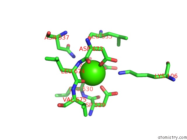
Calcium binding site 1 out of 4 in 1us1
Go back to
Calcium binding site 1 out
of 4 in the Crystal Structure of Human Vascular Adhesion Protein-1
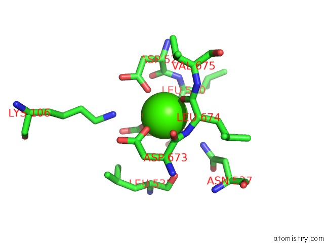
Mono view
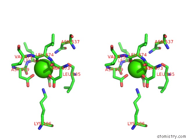
Stereo pair view
Mono view
Stereo pair view
A full contact list of Calcium with other atoms in the Ca binding
site number 1 of Crystal Structure of Human Vascular Adhesion Protein-1 within 5.0Å range:
|
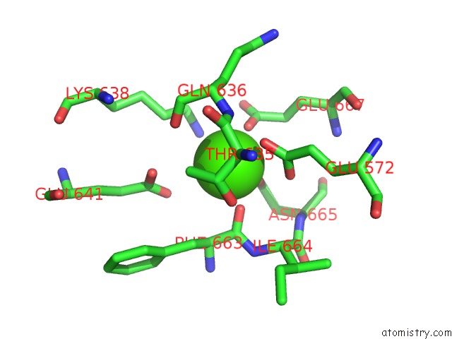
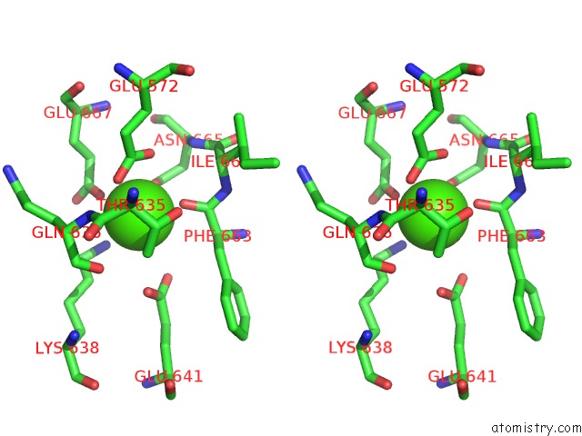
Calcium binding site 2 out of 4 in 1us1
Go back to
Calcium binding site 2 out
of 4 in the Crystal Structure of Human Vascular Adhesion Protein-1
Mono view
Stereo pair view
Mono view
Stereo pair view
A full contact list of Calcium with other atoms in the Ca binding
site number 2 of Crystal Structure of Human Vascular Adhesion Protein-1 within 5.0Å range:
|
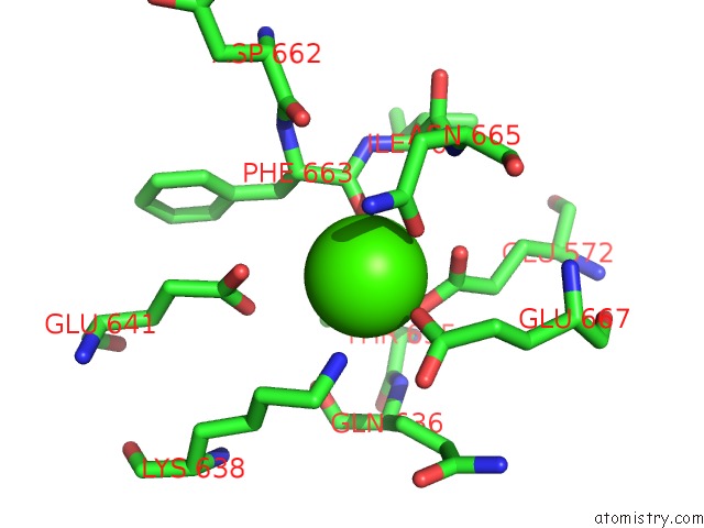
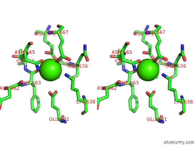
Calcium binding site 3 out of 4 in 1us1
Go back to
Calcium binding site 3 out
of 4 in the Crystal Structure of Human Vascular Adhesion Protein-1
Mono view

Stereo pair view
Mono view
Stereo pair view
A full contact list of Calcium with other atoms in the Ca binding
site number 3 of Crystal Structure of Human Vascular Adhesion Protein-1 within 5.0Å range:
|
Calcium binding site 4 out of 4 in 1us1
Go back to
Calcium binding site 4 out
of 4 in the Crystal Structure of Human Vascular Adhesion Protein-1
Mono view
Stereo pair view
Mono view
Stereo pair view
A full contact list of Calcium with other atoms in the Ca binding
site number 4 of Crystal Structure of Human Vascular Adhesion Protein-1 within 5.0Å range:
|
Reference:
T.T.Airenne,
Y.Nymalm,
H.Kidron,
D.J.Smith,
M.Pihlavisto,
M.Salmi,
S.Jalkanen,
M.S.Johnson,
T.A.Salminen.
Crystal Structure of the Human Vascular Adhesion Protein-1: Unique Structural Features with Functional Implications. Protein Sci. V. 14 1964 2005.
ISSN: ISSN 0961-8368
PubMed: 16046623
DOI: 10.1110/PS.051438105
Page generated: Thu Jul 11 23:37:43 2024
ISSN: ISSN 0961-8368
PubMed: 16046623
DOI: 10.1110/PS.051438105
Last articles
Zn in 9MJ5Zn in 9HNW
Zn in 9G0L
Zn in 9FNE
Zn in 9DZN
Zn in 9E0I
Zn in 9D32
Zn in 9DAK
Zn in 8ZXC
Zn in 8ZUF