Calcium »
PDB 1ux7-1v3d »
1uy3 »
Calcium in PDB 1uy3: Binding Sub-Site Dissection of A Family 6 Carbohydrate-Binding Module By X-Ray Crystallography and Isothermal Titration Calorimetry
Protein crystallography data
The structure of Binding Sub-Site Dissection of A Family 6 Carbohydrate-Binding Module By X-Ray Crystallography and Isothermal Titration Calorimetry, PDB code: 1uy3
was solved by
A.L.Van Bueren,
A.B.Boraston,
with X-Ray Crystallography technique. A brief refinement statistics is given in the table below:
| Resolution Low / High (Å) | 20.00 / 1.89 |
| Space group | P 41 21 2 |
| Cell size a, b, c (Å), α, β, γ (°) | 83.413, 83.413, 44.659, 90.00, 90.00, 90.00 |
| R / Rfree (%) | 13 / 16.6 |
Other elements in 1uy3:
The structure of Binding Sub-Site Dissection of A Family 6 Carbohydrate-Binding Module By X-Ray Crystallography and Isothermal Titration Calorimetry also contains other interesting chemical elements:
| Sodium | (Na) | 1 atom |
Calcium Binding Sites:
The binding sites of Calcium atom in the Binding Sub-Site Dissection of A Family 6 Carbohydrate-Binding Module By X-Ray Crystallography and Isothermal Titration Calorimetry
(pdb code 1uy3). This binding sites where shown within
5.0 Angstroms radius around Calcium atom.
In total only one binding site of Calcium was determined in the Binding Sub-Site Dissection of A Family 6 Carbohydrate-Binding Module By X-Ray Crystallography and Isothermal Titration Calorimetry, PDB code: 1uy3:
In total only one binding site of Calcium was determined in the Binding Sub-Site Dissection of A Family 6 Carbohydrate-Binding Module By X-Ray Crystallography and Isothermal Titration Calorimetry, PDB code: 1uy3:
Calcium binding site 1 out of 1 in 1uy3
Go back to
Calcium binding site 1 out
of 1 in the Binding Sub-Site Dissection of A Family 6 Carbohydrate-Binding Module By X-Ray Crystallography and Isothermal Titration Calorimetry
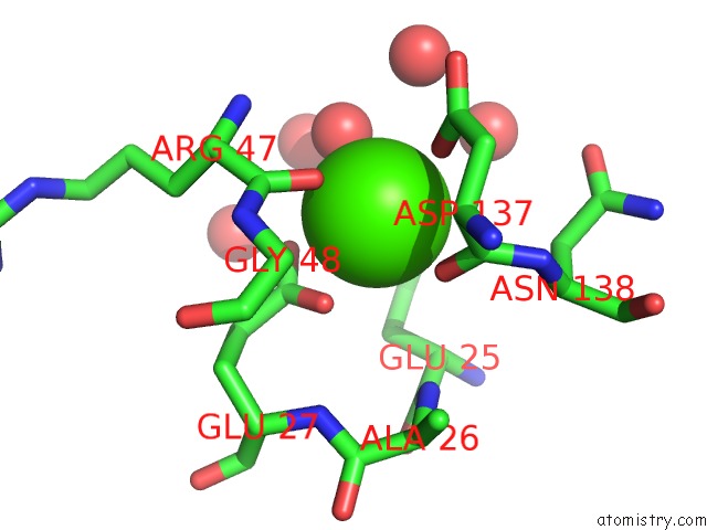
Mono view
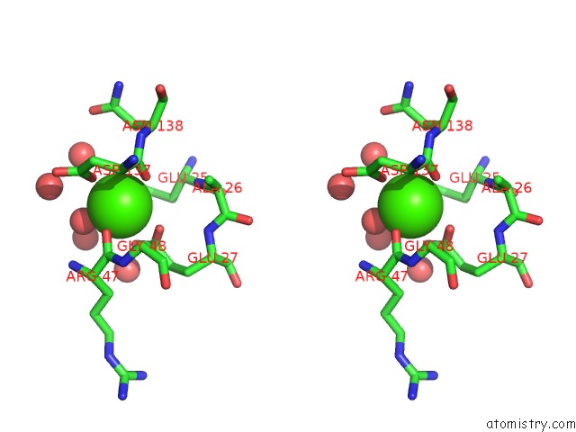
Stereo pair view
Mono view
Stereo pair view
A full contact list of Calcium with other atoms in the Ca binding
site number 1 of Binding Sub-Site Dissection of A Family 6 Carbohydrate-Binding Module By X-Ray Crystallography and Isothermal Titration Calorimetry within 5.0Å range:
|
Reference:
A.L.Van Bueren,
A.B.Boraston.
Binding Sub-Site Dissection of A Carbohydrate-Binding Module Reveals the Contribution of Entropy to Oligosaccharide Recognition at "Non-Primary" Binding Subsites. J.Mol.Biol. V. 340 869 2004.
ISSN: ISSN 0022-2836
PubMed: 15223327
DOI: 10.1016/J.JMB.2004.05.038
Page generated: Thu Jul 11 23:44:57 2024
ISSN: ISSN 0022-2836
PubMed: 15223327
DOI: 10.1016/J.JMB.2004.05.038
Last articles
Zn in 9JYWZn in 9IR4
Zn in 9IR3
Zn in 9GMX
Zn in 9GMW
Zn in 9JEJ
Zn in 9ERF
Zn in 9ERE
Zn in 9EGV
Zn in 9EGW