Calcium »
PDB 1v3e-1vfm »
1v3e »
Calcium in PDB 1v3e: Structure of the Hemagglutinin-Neuraminidase From Human Parainfluenza Virus Type III: Complex with Zanamavir
Enzymatic activity of Structure of the Hemagglutinin-Neuraminidase From Human Parainfluenza Virus Type III: Complex with Zanamavir
All present enzymatic activity of Structure of the Hemagglutinin-Neuraminidase From Human Parainfluenza Virus Type III: Complex with Zanamavir:
3.2.1.18;
3.2.1.18;
Protein crystallography data
The structure of Structure of the Hemagglutinin-Neuraminidase From Human Parainfluenza Virus Type III: Complex with Zanamavir, PDB code: 1v3e
was solved by
M.C.Lawrence,
N.A.Borg,
V.A.Streltsov,
P.A.Pilling,
V.C.Epa,
J.N.Varghese,
J.L.Mckimm-Breschkin,
P.M.Colman,
with X-Ray Crystallography technique. A brief refinement statistics is given in the table below:
| Resolution Low / High (Å) | 29.97 / 1.89 |
| Space group | P 62 2 2 |
| Cell size a, b, c (Å), α, β, γ (°) | 218.451, 218.451, 109.904, 90.00, 90.00, 120.00 |
| R / Rfree (%) | 18.4 / 21.9 |
Calcium Binding Sites:
The binding sites of Calcium atom in the Structure of the Hemagglutinin-Neuraminidase From Human Parainfluenza Virus Type III: Complex with Zanamavir
(pdb code 1v3e). This binding sites where shown within
5.0 Angstroms radius around Calcium atom.
In total 2 binding sites of Calcium where determined in the Structure of the Hemagglutinin-Neuraminidase From Human Parainfluenza Virus Type III: Complex with Zanamavir, PDB code: 1v3e:
Jump to Calcium binding site number: 1; 2;
In total 2 binding sites of Calcium where determined in the Structure of the Hemagglutinin-Neuraminidase From Human Parainfluenza Virus Type III: Complex with Zanamavir, PDB code: 1v3e:
Jump to Calcium binding site number: 1; 2;
Calcium binding site 1 out of 2 in 1v3e
Go back to
Calcium binding site 1 out
of 2 in the Structure of the Hemagglutinin-Neuraminidase From Human Parainfluenza Virus Type III: Complex with Zanamavir
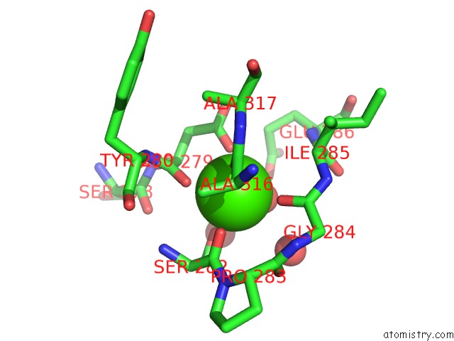
Mono view
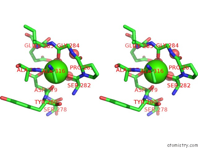
Stereo pair view
Mono view
Stereo pair view
A full contact list of Calcium with other atoms in the Ca binding
site number 1 of Structure of the Hemagglutinin-Neuraminidase From Human Parainfluenza Virus Type III: Complex with Zanamavir within 5.0Å range:
|
Calcium binding site 2 out of 2 in 1v3e
Go back to
Calcium binding site 2 out
of 2 in the Structure of the Hemagglutinin-Neuraminidase From Human Parainfluenza Virus Type III: Complex with Zanamavir
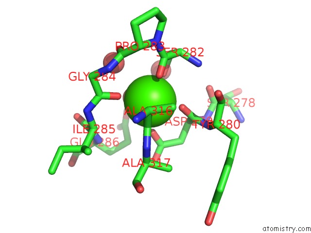
Mono view
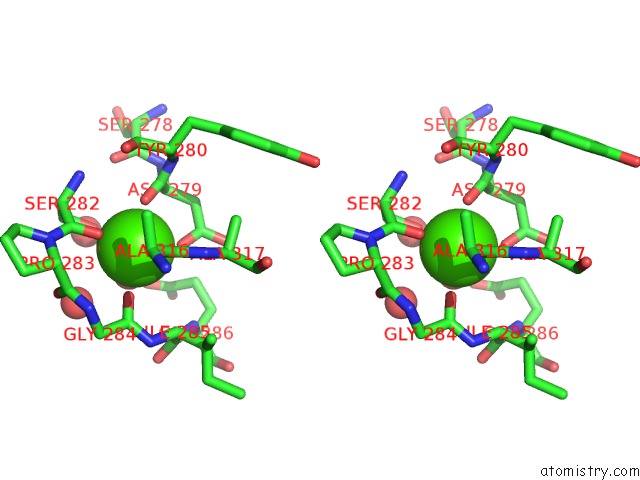
Stereo pair view
Mono view
Stereo pair view
A full contact list of Calcium with other atoms in the Ca binding
site number 2 of Structure of the Hemagglutinin-Neuraminidase From Human Parainfluenza Virus Type III: Complex with Zanamavir within 5.0Å range:
|
Reference:
M.C.Lawrence,
N.A.Borg,
V.A.Streltsov,
P.A.Pilling,
V.C.Epa,
J.N.Varghese,
J.L.Mckimm-Breschkin,
P.M.Colman.
Structure of the Haemagglutinin-Neuraminidase From Human Parainfluenza Virus Type III J.Mol.Biol. V. 335 1343 2004.
ISSN: ISSN 0022-2836
PubMed: 14729348
DOI: 10.1016/J.JMB.2003.11.032
Page generated: Tue Jul 8 02:51:19 2025
ISSN: ISSN 0022-2836
PubMed: 14729348
DOI: 10.1016/J.JMB.2003.11.032
Last articles
Fe in 2YXOFe in 2YRS
Fe in 2YXC
Fe in 2YNM
Fe in 2YVJ
Fe in 2YP1
Fe in 2YU2
Fe in 2YU1
Fe in 2YQB
Fe in 2YOO