Atomistry » Calcium » PDB 1v3e-1vfm » 1v3k
Atomistry »
  Calcium »
    PDB 1v3e-1vfm »
      1v3k »

Calcium in PDB 1v3k: Crystal Structure of F283Y Mutant Cyclodextrin Glycosyltransferase

Enzymatic activity of Crystal Structure of F283Y Mutant Cyclodextrin Glycosyltransferase

All present enzymatic activity of Crystal Structure of F283Y Mutant Cyclodextrin Glycosyltransferase:
2.4.1.19;

Protein crystallography data

The structure of Crystal Structure of F283Y Mutant Cyclodextrin Glycosyltransferase, PDB code: 1v3k was solved by R.Kanai, K.Haga, T.Akiba, K.Yamane, K.Harata, with X-Ray Crystallography technique. A brief refinement statistics is given in the table below:

Resolution Low / High (Å) 10.00 / 2.00
Space group P 1
Cell size a, b, c (Å), α, β, γ (°) 64.880, 74.430, 79.010, 85.15, 105.05, 101.02
R / Rfree (%) 15.5 / 20

Calcium Binding Sites:

The binding sites of Calcium atom in the Crystal Structure of F283Y Mutant Cyclodextrin Glycosyltransferase (pdb code 1v3k). This binding sites where shown within 5.0 Angstroms radius around Calcium atom.
In total 4 binding sites of Calcium where determined in the Crystal Structure of F283Y Mutant Cyclodextrin Glycosyltransferase, PDB code: 1v3k:
Jump to Calcium binding site number: 1; 2; 3; 4;

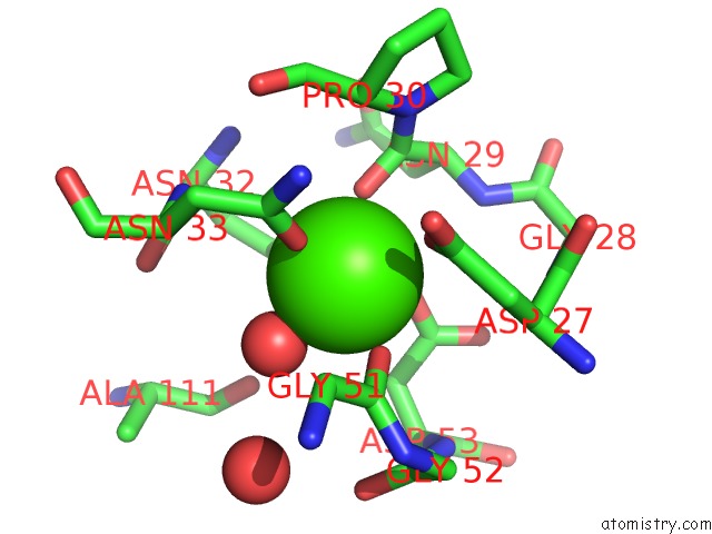
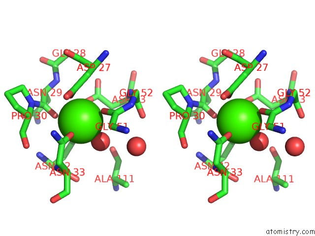
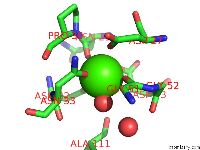
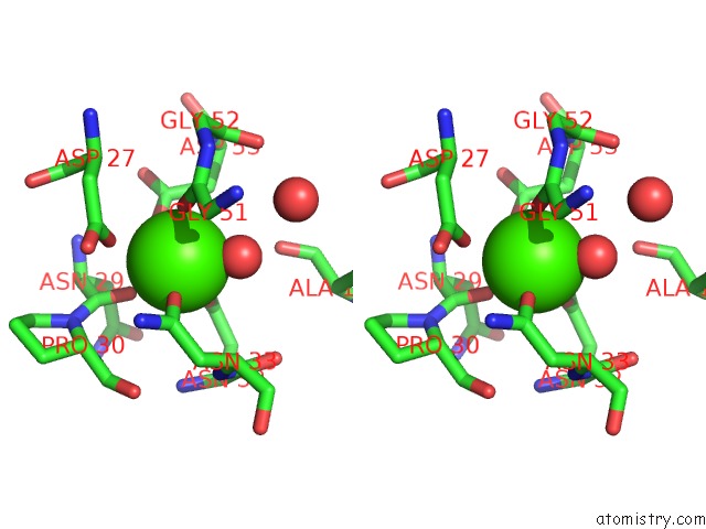
Calcium binding site 1 out of 4 in 1v3k

Go back to Calcium Binding Sites List in 1v3k
Calcium binding site 1 out of 4 in the Crystal Structure of F283Y Mutant Cyclodextrin Glycosyltransferase


Mono view


Stereo pair view

A full contact list of Calcium with other atoms in the Ca binding site number 1 of Crystal Structure of F283Y Mutant Cyclodextrin Glycosyltransferase within 5.0Å range:
probe atom residue distance (Å) B Occ
A:Ca687

b:7.2
occ:1.00
OD1 A:ASN33 2.1 11.9 1.0
OD1 A:ASN32 2.2 10.4 1.0
O A:HOH1746 2.3 7.5 1.0
O A:ASN29 2.3 11.6 1.0
O A:GLY51 2.3 13.5 1.0
OD2 A:ASP53 2.4 11.2 1.0
OD2 A:ASP27 2.4 9.0 1.0
CG A:ASP27 3.3 10.3 1.0
CG A:ASN33 3.4 10.4 1.0
C A:ASN29 3.4 11.5 1.0
CG A:ASN32 3.4 9.6 1.0
CG A:ASP53 3.5 10.4 1.0
C A:GLY51 3.5 10.9 1.0
OD1 A:ASP27 3.9 9.3 1.0
CB A:ASP53 4.0 8.6 1.0
CA A:GLY51 4.1 8.0 1.0
N A:PRO30 4.1 13.8 1.0
N A:ASN33 4.1 9.7 1.0
ND2 A:ASN32 4.1 6.9 1.0
ND2 A:ASN33 4.2 9.0 1.0
CA A:PRO30 4.2 15.5 1.0
N A:ASN29 4.3 10.1 1.0
O A:ALA111 4.3 10.6 1.0
CA A:ASN33 4.3 9.8 1.0
CB A:ASP27 4.3 7.0 1.0
CA A:ASN29 4.4 11.7 1.0
C A:ASN32 4.4 9.6 1.0
CB A:ASN33 4.4 10.3 1.0
OD1 A:ASP53 4.4 8.2 1.0
CA A:ASP27 4.5 8.9 1.0
N A:GLY52 4.6 9.6 1.0
CB A:ASN32 4.7 10.3 1.0
C A:PRO30 4.7 14.2 1.0
N A:ASN32 4.7 12.1 1.0
C A:GLY52 4.7 8.2 1.0
O A:PRO30 4.8 16.9 1.0
CB A:ASN29 4.8 11.3 1.0
O A:ASN32 4.8 9.2 1.0
CA A:ASN32 4.8 10.1 1.0
N A:ASP53 4.8 9.1 1.0
O A:HOH1082 4.9 14.8 1.0
C A:ASP27 4.9 10.7 1.0
O A:GLY52 5.0 7.6 1.0
CA A:GLY52 5.0 9.9 1.0
N A:GLY28 5.0 10.9 1.0

Calcium binding site 2 out of 4 in 1v3k

Go back to Calcium Binding Sites List in 1v3k
Calcium binding site 2 out of 4 in the Crystal Structure of F283Y Mutant Cyclodextrin Glycosyltransferase


Mono view


Stereo pair view

A full contact list of Calcium with other atoms in the Ca binding site number 2 of Crystal Structure of F283Y Mutant Cyclodextrin Glycosyltransferase within 5.0Å range:
probe atom residue distance (Å) B Occ
A:Ca688

b:11.8
occ:1.00
O A:HIS233 2.3 4.5 1.0
OD1 A:ASN139 2.3 4.5 1.0
O A:HOH1024 2.3 7.9 1.0
O A:HOH1759 2.3 9.4 1.0
O A:ILE190 2.3 6.9 1.0
O A:HOH1134 2.4 12.9 1.0
OD1 A:ASP199 2.6 14.3 1.0
OD2 A:ASP199 2.9 14.5 1.0
CG A:ASP199 3.1 13.9 1.0
CG A:ASN139 3.4 6.0 1.0
C A:ILE190 3.5 8.7 1.0
C A:HIS233 3.5 5.1 1.0
ND2 A:ASN139 3.9 7.5 1.0
CA A:ILE190 4.1 7.2 1.0
O A:ASN139 4.4 9.1 1.0
N A:MET234 4.4 6.5 1.0
CA A:MET234 4.4 7.2 1.0
CB A:HIS233 4.5 5.0 1.0
CA A:HIS233 4.5 2.8 1.0
CG A:MET234 4.6 5.8 1.0
O A:GLY189 4.6 7.5 1.0
O A:LYS192 4.6 14.5 1.0
CB A:ASP199 4.6 10.0 1.0
N A:TYR191 4.6 8.1 1.0
CB A:ASN139 4.7 3.8 1.0
CG2 A:ILE190 4.7 6.0 1.0
O A:HOH1553 4.8 14.0 1.0
ND1 A:HIS176 4.8 10.6 1.0
O A:LEU200 4.9 9.0 1.0
CE1 A:HIS176 4.9 12.9 1.0
CA A:TYR191 5.0 8.3 1.0

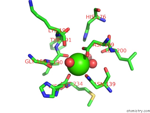
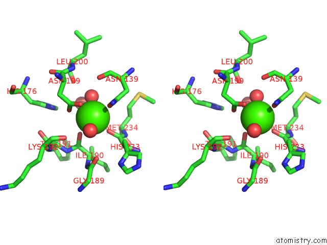
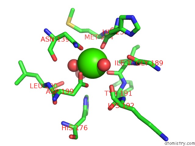
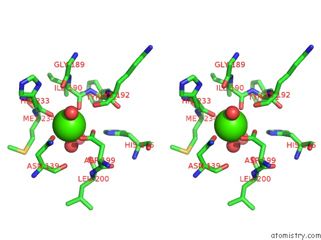
Calcium binding site 3 out of 4 in 1v3k

Go back to Calcium Binding Sites List in 1v3k
Calcium binding site 3 out of 4 in the Crystal Structure of F283Y Mutant Cyclodextrin Glycosyltransferase


Mono view


Stereo pair view

A full contact list of Calcium with other atoms in the Ca binding site number 3 of Crystal Structure of F283Y Mutant Cyclodextrin Glycosyltransferase within 5.0Å range:
probe atom residue distance (Å) B Occ
B:Ca689

b:13.3
occ:1.00
OD1 B:ASN33 2.1 13.8 1.0
OD1 B:ASN32 2.1 15.3 1.0
O B:HOH1783 2.2 15.7 1.0
O B:GLY51 2.3 12.4 1.0
O B:ASN29 2.3 19.1 1.0
OD2 B:ASP53 2.4 11.3 1.0
OD2 B:ASP27 2.7 14.7 1.0
CG B:ASN33 3.4 13.9 1.0
CG B:ASN32 3.4 14.8 1.0
CG B:ASP53 3.4 14.5 1.0
C B:GLY51 3.4 9.8 1.0
C B:ASN29 3.5 16.9 1.0
CG B:ASP27 3.6 14.1 1.0
CB B:ASP53 3.8 10.7 1.0
CA B:GLY51 4.0 9.7 1.0
ND2 B:ASN32 4.1 15.5 1.0
O B:ALA111 4.1 13.0 1.0
N B:ASN33 4.1 12.9 1.0
ND2 B:ASN33 4.2 12.8 1.0
OD1 B:ASP27 4.3 13.3 1.0
CA B:ASN33 4.3 14.2 1.0
N B:PRO30 4.3 18.7 1.0
N B:ASN29 4.4 17.1 1.0
C B:ASN32 4.4 13.5 1.0
CA B:PRO30 4.4 20.1 1.0
CB B:ASN33 4.4 14.4 1.0
CA B:ASN29 4.4 16.4 1.0
OD1 B:ASP53 4.5 13.8 1.0
CB B:ASP27 4.6 12.3 1.0
N B:GLY52 4.6 9.4 1.0
CB B:ASN32 4.6 14.9 1.0
CA B:ASP27 4.6 12.9 1.0
C B:GLY52 4.7 10.2 1.0
O B:HOH1782 4.7 18.3 1.0
N B:ASN32 4.7 17.0 1.0
CB B:ASN29 4.7 16.9 1.0
C B:PRO30 4.7 19.5 1.0
O B:ASN32 4.8 15.1 1.0
O B:PRO30 4.8 21.3 1.0
CA B:ASN32 4.8 15.0 1.0
N B:ASP53 4.8 7.7 1.0
O B:GLY52 4.9 9.4 1.0
CA B:ASP53 5.0 8.2 1.0
CA B:GLY52 5.0 9.7 1.0

Calcium binding site 4 out of 4 in 1v3k

Go back to Calcium Binding Sites List in 1v3k
Calcium binding site 4 out of 4 in the Crystal Structure of F283Y Mutant Cyclodextrin Glycosyltransferase


Mono view


Stereo pair view

A full contact list of Calcium with other atoms in the Ca binding site number 4 of Crystal Structure of F283Y Mutant Cyclodextrin Glycosyltransferase within 5.0Å range:
probe atom residue distance (Å) B Occ
B:Ca690

b:13.1
occ:1.00
O B:HOH1120 2.2 11.8 1.0
O B:HIS233 2.3 9.9 1.0
O B:HOH1052 2.3 8.9 1.0
O B:ILE190 2.3 8.7 1.0
OD1 B:ASN139 2.3 11.1 1.0
O B:HOH1563 2.3 14.7 1.0
OD2 B:ASP199 2.7 15.5 1.0
OD1 B:ASP199 2.7 14.4 1.0
CG B:ASP199 3.0 15.8 1.0
C B:ILE190 3.5 10.0 1.0
CG B:ASN139 3.5 10.0 1.0
C B:HIS233 3.6 11.0 1.0
ND2 B:ASN139 4.0 12.5 1.0
CA B:ILE190 4.2 10.9 1.0
O B:LYS192 4.4 14.5 1.0
CB B:HIS233 4.4 9.8 1.0
N B:MET234 4.4 9.0 1.0
O B:ASN139 4.4 9.5 1.0
CA B:MET234 4.5 9.3 1.0
CA B:HIS233 4.5 11.3 1.0
O B:GLY189 4.5 12.9 1.0
CB B:ASP199 4.6 10.6 1.0
N B:TYR191 4.6 10.5 1.0
CG B:MET234 4.7 8.2 1.0
O B:HOH1576 4.7 23.8 1.0
CB B:ASN139 4.8 9.7 1.0
CG2 B:ILE190 4.8 11.1 1.0
ND1 B:HIS176 4.8 14.3 1.0
O B:LEU200 4.9 11.2 1.0
CA B:TYR191 4.9 12.0 1.0

Reference:

R.Kanai, K.Haga, T.Akiba, K.Yamane, K.Harata. Role of PHE283 in Enzymatic Reaction of Cyclodextrin Glycosyltransferase From Alkalophilic Bacillus Sp.1011: Substrate Binding and Arrangement of the Catalytic Site Protein Sci. V. 13 457 2004.
ISSN: ISSN 0961-8368
PubMed: 14739329
DOI: 10.1110/PS.03408504
Page generated: Sat Dec 12 03:24:25 2020

Last articles

Zn in 8WB0
Zn in 8WAX
Zn in 8WAU
Zn in 8WAZ
Zn in 8WAY
Zn in 8WAV
Zn in 8WAW
Zn in 8WAT
Zn in 8W7M
Zn in 8WD3
© Copyright 2008-2020 by atomistry.com
Home   |    Site Map   |    Copyright   |    Contact us   |    Privacy