Calcium »
PDB 1v3e-1vfm »
1v6o »
Calcium in PDB 1v6o: Peanut Lectin Complexed with 10MER Peptide (Pvriwssatg)
Protein crystallography data
The structure of Peanut Lectin Complexed with 10MER Peptide (Pvriwssatg), PDB code: 1v6o
was solved by
S.Kundhavai Natchiar,
A.Arockia Jeyaprakash,
T.N.C.Ramya,
C.J.Thomas,
K.Suguna,
A.Surolia,
M.Vijayan,
with X-Ray Crystallography technique. A brief refinement statistics is given in the table below:
| Resolution Low / High (Å) | 20.00 / 3.00 |
| Space group | P 1 21 1 |
| Cell size a, b, c (Å), α, β, γ (°) | 128.127, 125.800, 84.740, 90.00, 116.19, 90.00 |
| R / Rfree (%) | 17.4 / 23.9 |
Other elements in 1v6o:
The structure of Peanut Lectin Complexed with 10MER Peptide (Pvriwssatg) also contains other interesting chemical elements:
| Manganese | (Mn) | 8 atoms |
Calcium Binding Sites:
The binding sites of Calcium atom in the Peanut Lectin Complexed with 10MER Peptide (Pvriwssatg)
(pdb code 1v6o). This binding sites where shown within
5.0 Angstroms radius around Calcium atom.
In total 8 binding sites of Calcium where determined in the Peanut Lectin Complexed with 10MER Peptide (Pvriwssatg), PDB code: 1v6o:
Jump to Calcium binding site number: 1; 2; 3; 4; 5; 6; 7; 8;
In total 8 binding sites of Calcium where determined in the Peanut Lectin Complexed with 10MER Peptide (Pvriwssatg), PDB code: 1v6o:
Jump to Calcium binding site number: 1; 2; 3; 4; 5; 6; 7; 8;
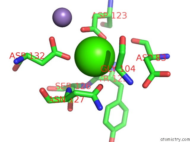
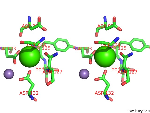
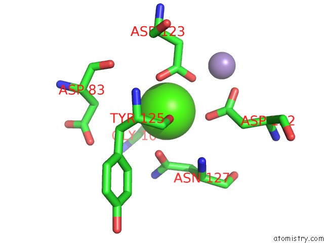
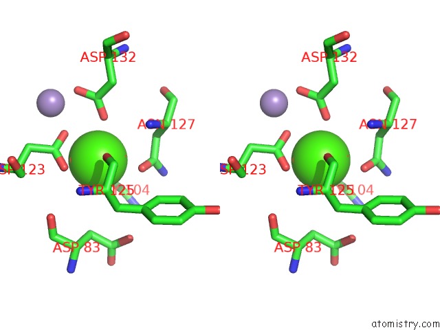
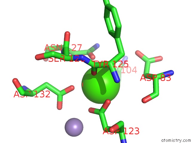
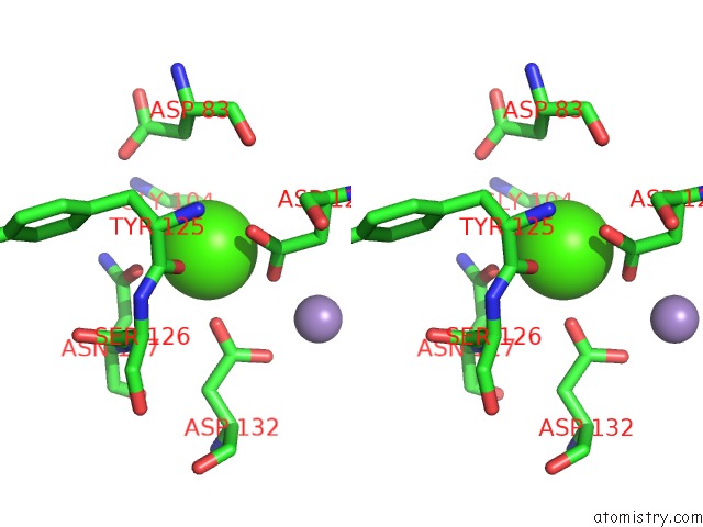
Calcium binding site 1 out of 8 in 1v6o
Go back to
Calcium binding site 1 out
of 8 in the Peanut Lectin Complexed with 10MER Peptide (Pvriwssatg)
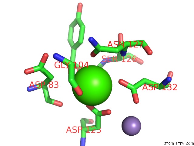
Mono view
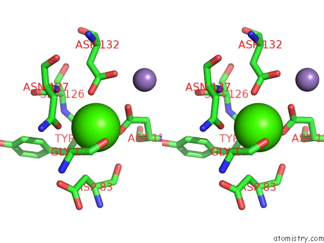
Stereo pair view
Mono view
Stereo pair view
A full contact list of Calcium with other atoms in the Ca binding
site number 1 of Peanut Lectin Complexed with 10MER Peptide (Pvriwssatg) within 5.0Å range:
|
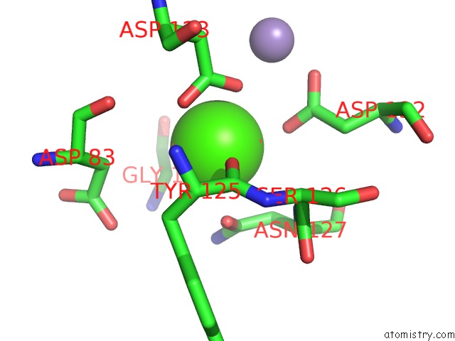
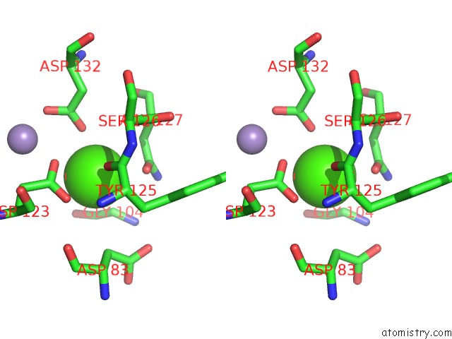
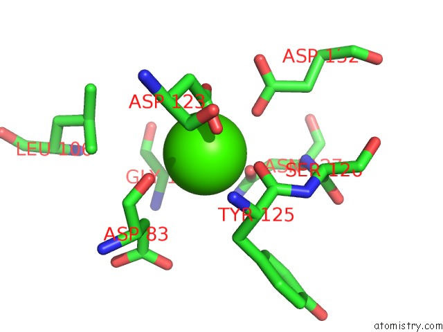
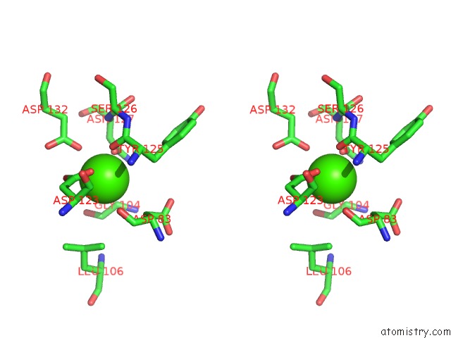
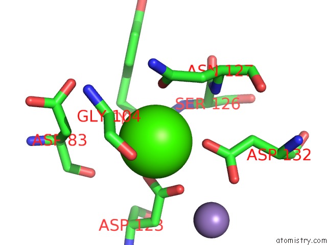
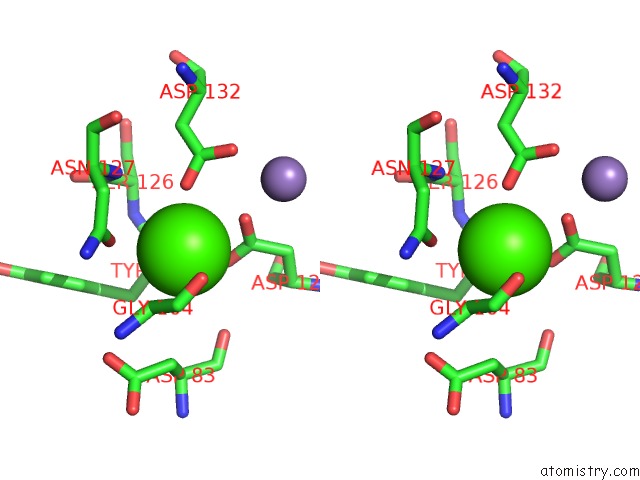
Calcium binding site 2 out of 8 in 1v6o
Go back to
Calcium binding site 2 out
of 8 in the Peanut Lectin Complexed with 10MER Peptide (Pvriwssatg)
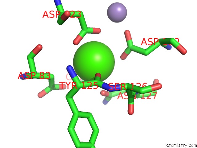
Mono view
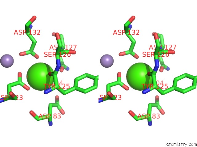
Stereo pair view
Mono view
Stereo pair view
A full contact list of Calcium with other atoms in the Ca binding
site number 2 of Peanut Lectin Complexed with 10MER Peptide (Pvriwssatg) within 5.0Å range:
|
Calcium binding site 3 out of 8 in 1v6o
Go back to
Calcium binding site 3 out
of 8 in the Peanut Lectin Complexed with 10MER Peptide (Pvriwssatg)
Mono view
Stereo pair view
Mono view
Stereo pair view
A full contact list of Calcium with other atoms in the Ca binding
site number 3 of Peanut Lectin Complexed with 10MER Peptide (Pvriwssatg) within 5.0Å range:
|
Calcium binding site 4 out of 8 in 1v6o
Go back to
Calcium binding site 4 out
of 8 in the Peanut Lectin Complexed with 10MER Peptide (Pvriwssatg)
Mono view
Stereo pair view
Mono view
Stereo pair view
A full contact list of Calcium with other atoms in the Ca binding
site number 4 of Peanut Lectin Complexed with 10MER Peptide (Pvriwssatg) within 5.0Å range:
|
Calcium binding site 5 out of 8 in 1v6o
Go back to
Calcium binding site 5 out
of 8 in the Peanut Lectin Complexed with 10MER Peptide (Pvriwssatg)
Mono view
Stereo pair view
Mono view
Stereo pair view
A full contact list of Calcium with other atoms in the Ca binding
site number 5 of Peanut Lectin Complexed with 10MER Peptide (Pvriwssatg) within 5.0Å range:
|
Calcium binding site 6 out of 8 in 1v6o
Go back to
Calcium binding site 6 out
of 8 in the Peanut Lectin Complexed with 10MER Peptide (Pvriwssatg)
Mono view
Stereo pair view
Mono view
Stereo pair view
A full contact list of Calcium with other atoms in the Ca binding
site number 6 of Peanut Lectin Complexed with 10MER Peptide (Pvriwssatg) within 5.0Å range:
|
Calcium binding site 7 out of 8 in 1v6o
Go back to
Calcium binding site 7 out
of 8 in the Peanut Lectin Complexed with 10MER Peptide (Pvriwssatg)
Mono view
Stereo pair view
Mono view
Stereo pair view
A full contact list of Calcium with other atoms in the Ca binding
site number 7 of Peanut Lectin Complexed with 10MER Peptide (Pvriwssatg) within 5.0Å range:
|
Calcium binding site 8 out of 8 in 1v6o
Go back to
Calcium binding site 8 out
of 8 in the Peanut Lectin Complexed with 10MER Peptide (Pvriwssatg)
Mono view
Stereo pair view
Mono view
Stereo pair view
A full contact list of Calcium with other atoms in the Ca binding
site number 8 of Peanut Lectin Complexed with 10MER Peptide (Pvriwssatg) within 5.0Å range:
|
Reference:
S.Kundhavai Natchiar,
A.Arockia Jeyaprakash,
T.N.Ramya,
C.J.Thomas,
K.Suguna,
A.Surolia,
M.Vijayan.
Structural Plasticity of Peanut Lectin: An X-Ray Analysis Involving Variation in pH, Ligand Binding and Crystal Structure. Acta Crystallogr.,Sect.D V. 60 211 2004.
ISSN: ISSN 0907-4449
PubMed: 14747696
DOI: 10.1107/S090744490302849X
Page generated: Thu Jul 11 23:58:31 2024
ISSN: ISSN 0907-4449
PubMed: 14747696
DOI: 10.1107/S090744490302849X
Last articles
Zn in 9JYWZn in 9IR4
Zn in 9IR3
Zn in 9GMX
Zn in 9GMW
Zn in 9JEJ
Zn in 9ERF
Zn in 9ERE
Zn in 9EGV
Zn in 9EGW