Calcium »
PDB 1v3e-1vfm »
1v7v »
Calcium in PDB 1v7v: Crystal Structure of Vibrio Proteolyticus Chitobiose Phosphorylase
Protein crystallography data
The structure of Crystal Structure of Vibrio Proteolyticus Chitobiose Phosphorylase, PDB code: 1v7v
was solved by
M.Hidaka,
Y.Honda,
S.Nirasawa,
M.Kitaoka,
K.Hayashi,
T.Wakagi,
H.Shoun,
S.Fushinobu,
with X-Ray Crystallography technique. A brief refinement statistics is given in the table below:
| Resolution Low / High (Å) | 47.78 / 1.80 |
| Space group | C 1 2 1 |
| Cell size a, b, c (Å), α, β, γ (°) | 140.946, 70.580, 80.045, 90.00, 98.40, 90.00 |
| R / Rfree (%) | 16.2 / 18.6 |
Calcium Binding Sites:
The binding sites of Calcium atom in the Crystal Structure of Vibrio Proteolyticus Chitobiose Phosphorylase
(pdb code 1v7v). This binding sites where shown within
5.0 Angstroms radius around Calcium atom.
In total 3 binding sites of Calcium where determined in the Crystal Structure of Vibrio Proteolyticus Chitobiose Phosphorylase, PDB code: 1v7v:
Jump to Calcium binding site number: 1; 2; 3;
In total 3 binding sites of Calcium where determined in the Crystal Structure of Vibrio Proteolyticus Chitobiose Phosphorylase, PDB code: 1v7v:
Jump to Calcium binding site number: 1; 2; 3;
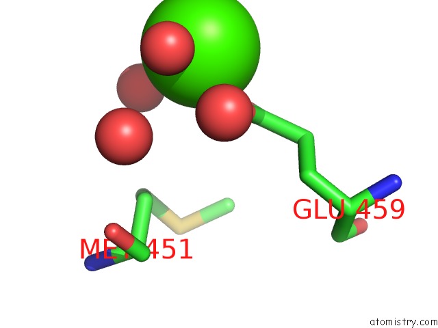
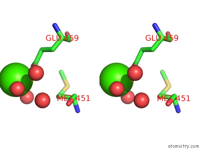
Calcium binding site 1 out of 3 in 1v7v
Go back to
Calcium binding site 1 out
of 3 in the Crystal Structure of Vibrio Proteolyticus Chitobiose Phosphorylase
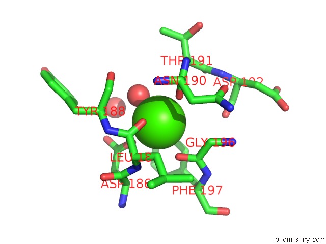
Mono view
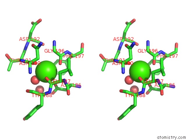
Stereo pair view
Mono view
Stereo pair view
A full contact list of Calcium with other atoms in the Ca binding
site number 1 of Crystal Structure of Vibrio Proteolyticus Chitobiose Phosphorylase within 5.0Å range:
|
Calcium binding site 2 out of 3 in 1v7v
Go back to
Calcium binding site 2 out
of 3 in the Crystal Structure of Vibrio Proteolyticus Chitobiose Phosphorylase
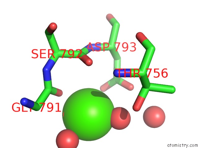
Mono view
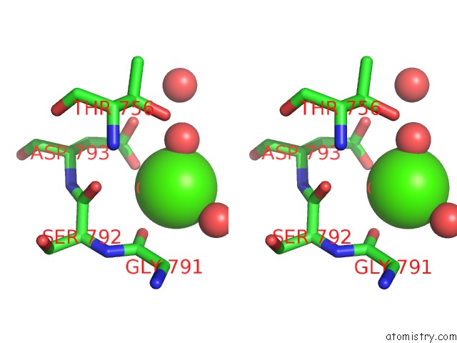
Stereo pair view
Mono view
Stereo pair view
A full contact list of Calcium with other atoms in the Ca binding
site number 2 of Crystal Structure of Vibrio Proteolyticus Chitobiose Phosphorylase within 5.0Å range:
|
Calcium binding site 3 out of 3 in 1v7v
Go back to
Calcium binding site 3 out
of 3 in the Crystal Structure of Vibrio Proteolyticus Chitobiose Phosphorylase
Mono view
Stereo pair view
Mono view
Stereo pair view
A full contact list of Calcium with other atoms in the Ca binding
site number 3 of Crystal Structure of Vibrio Proteolyticus Chitobiose Phosphorylase within 5.0Å range:
|
Reference:
M.Hidaka,
Y.Honda,
M.Kitaoka,
S.Nirasawa,
K.Hayashi,
T.Wakagi,
H.Shoun,
S.Fushinobu.
Chitobiose Phosphorylase From Vibrio Proteolyticus, A Member of Glycosyl Transferase Family 36, Has A Clan Gh-L-Like (Alpha/Alpha)(6) Barrel Fold. Structure V. 12 937 2004.
ISSN: ISSN 0969-2126
PubMed: 15274915
DOI: 10.1016/J.STR.2004.03.027
Page generated: Tue Jul 8 02:56:17 2025
ISSN: ISSN 0969-2126
PubMed: 15274915
DOI: 10.1016/J.STR.2004.03.027
Last articles
Cl in 8AY7Cl in 8AY3
Cl in 8AY6
Cl in 8AXT
Cl in 8AXS
Cl in 8AXW
Cl in 8AXU
Cl in 8AXI
Cl in 8AXC
Cl in 8AXR