Calcium »
PDB 1v3e-1vfm »
1v9u »
Calcium in PDB 1v9u: Human Rhinovirus 2 Bound to A Fragment of Its Cellular Receptor Protein
Protein crystallography data
The structure of Human Rhinovirus 2 Bound to A Fragment of Its Cellular Receptor Protein, PDB code: 1v9u
was solved by
N.Verdaguer,
I.Fita,
M.Reithmayer,
R.Moser,
D.Blaas,
with X-Ray Crystallography technique. A brief refinement statistics is given in the table below:
| Resolution Low / High (Å) | 20.00 / 3.60 |
| Space group | P 21 2 21 |
| Cell size a, b, c (Å), α, β, γ (°) | 313.100, 348.790, 380.890, 90.00, 90.00, 90.00 |
| R / Rfree (%) | 28.5 / 29.5 |
Calcium Binding Sites:
The binding sites of Calcium atom in the Human Rhinovirus 2 Bound to A Fragment of Its Cellular Receptor Protein
(pdb code 1v9u). This binding sites where shown within
5.0 Angstroms radius around Calcium atom.
In total only one binding site of Calcium was determined in the Human Rhinovirus 2 Bound to A Fragment of Its Cellular Receptor Protein, PDB code: 1v9u:
In total only one binding site of Calcium was determined in the Human Rhinovirus 2 Bound to A Fragment of Its Cellular Receptor Protein, PDB code: 1v9u:
Calcium binding site 1 out of 1 in 1v9u
Go back to
Calcium binding site 1 out
of 1 in the Human Rhinovirus 2 Bound to A Fragment of Its Cellular Receptor Protein
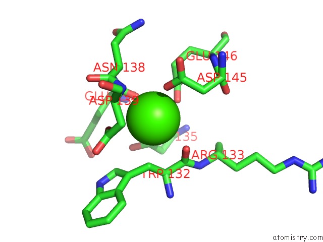
Mono view
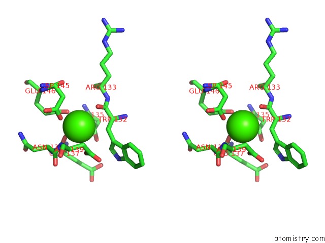
Stereo pair view
Mono view
Stereo pair view
A full contact list of Calcium with other atoms in the Ca binding
site number 1 of Human Rhinovirus 2 Bound to A Fragment of Its Cellular Receptor Protein within 5.0Å range:
|
Reference:
N.Verdaguer,
I.Fita,
M.Reithmayer,
R.Moser,
D.Blaas.
X-Ray Structure of A Minor Group Human Rhinovirus Bound to A Fragment of Its Cellular Receptor Protein Nat.Struct.Mol.Biol. V. 11 429 2004.
ISSN: ISSN 1545-9993
PubMed: 15064754
DOI: 10.1038/NSMB753
Page generated: Fri Jul 12 00:01:09 2024
ISSN: ISSN 1545-9993
PubMed: 15064754
DOI: 10.1038/NSMB753
Last articles
Zn in 9MJ5Zn in 9HNW
Zn in 9G0L
Zn in 9FNE
Zn in 9DZN
Zn in 9E0I
Zn in 9D32
Zn in 9DAK
Zn in 8ZXC
Zn in 8ZUF