Calcium »
PDB 1vfo-1w7c »
1vkq »
Calcium in PDB 1vkq: A Re-Determination of the Structure of the Triple Mutant (K53,56,120M) of Phospholipase A2 at 1.6A Resolution Using Sulphur-Sas at 1.54A Wavelength
Enzymatic activity of A Re-Determination of the Structure of the Triple Mutant (K53,56,120M) of Phospholipase A2 at 1.6A Resolution Using Sulphur-Sas at 1.54A Wavelength
All present enzymatic activity of A Re-Determination of the Structure of the Triple Mutant (K53,56,120M) of Phospholipase A2 at 1.6A Resolution Using Sulphur-Sas at 1.54A Wavelength:
3.1.1.4;
3.1.1.4;
Protein crystallography data
The structure of A Re-Determination of the Structure of the Triple Mutant (K53,56,120M) of Phospholipase A2 at 1.6A Resolution Using Sulphur-Sas at 1.54A Wavelength, PDB code: 1vkq
was solved by
K.Sekar,
D.Velmurugan,
V.Rajakannan,
T.Yamane,
M.Dauter,
Z.Dauter,
with X-Ray Crystallography technique. A brief refinement statistics is given in the table below:
| Resolution Low / High (Å) | 19.96 / 1.60 |
| Space group | P 31 2 1 |
| Cell size a, b, c (Å), α, β, γ (°) | 46.050, 46.050, 101.580, 90.00, 90.00, 120.00 |
| R / Rfree (%) | 17.7 / 21.7 |
Other elements in 1vkq:
The structure of A Re-Determination of the Structure of the Triple Mutant (K53,56,120M) of Phospholipase A2 at 1.6A Resolution Using Sulphur-Sas at 1.54A Wavelength also contains other interesting chemical elements:
| Chlorine | (Cl) | 1 atom |
Calcium Binding Sites:
The binding sites of Calcium atom in the A Re-Determination of the Structure of the Triple Mutant (K53,56,120M) of Phospholipase A2 at 1.6A Resolution Using Sulphur-Sas at 1.54A Wavelength
(pdb code 1vkq). This binding sites where shown within
5.0 Angstroms radius around Calcium atom.
In total only one binding site of Calcium was determined in the A Re-Determination of the Structure of the Triple Mutant (K53,56,120M) of Phospholipase A2 at 1.6A Resolution Using Sulphur-Sas at 1.54A Wavelength, PDB code: 1vkq:
In total only one binding site of Calcium was determined in the A Re-Determination of the Structure of the Triple Mutant (K53,56,120M) of Phospholipase A2 at 1.6A Resolution Using Sulphur-Sas at 1.54A Wavelength, PDB code: 1vkq:
Calcium binding site 1 out of 1 in 1vkq
Go back to
Calcium binding site 1 out
of 1 in the A Re-Determination of the Structure of the Triple Mutant (K53,56,120M) of Phospholipase A2 at 1.6A Resolution Using Sulphur-Sas at 1.54A Wavelength
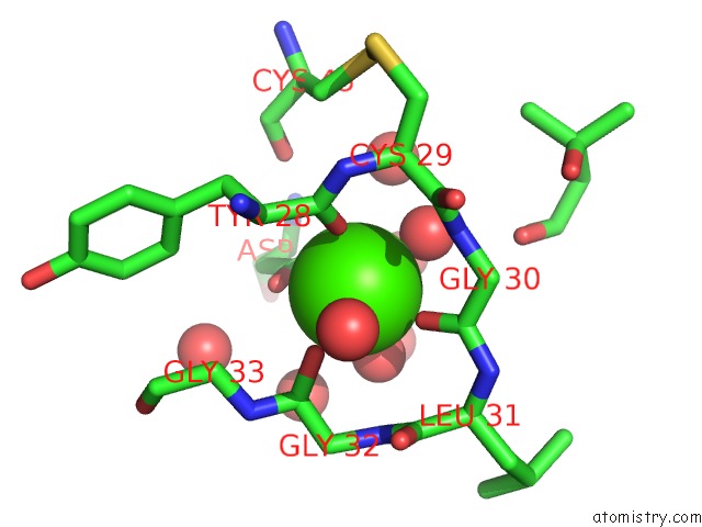
Mono view
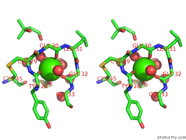
Stereo pair view
Mono view
Stereo pair view
A full contact list of Calcium with other atoms in the Ca binding
site number 1 of A Re-Determination of the Structure of the Triple Mutant (K53,56,120M) of Phospholipase A2 at 1.6A Resolution Using Sulphur-Sas at 1.54A Wavelength within 5.0Å range:
|
Reference:
K.Sekar,
V.Rajakannan,
D.Velmurugan,
T.Yamane,
R.Thirumurugan,
M.Dauter,
Z.Dauter.
A Redetermination of the Structure of the Triple Mutant (K53,56,120M) of Phospholipase A2 at 1.6 A Resolution Using Sulfur-Sas at 1.54 A Wavelength. Acta Crystallogr.,Sect.D V. 60 1586 2004.
ISSN: ISSN 0907-4449
PubMed: 15333929
DOI: 10.1107/S090744490401697X
Page generated: Fri Jul 12 06:55:33 2024
ISSN: ISSN 0907-4449
PubMed: 15333929
DOI: 10.1107/S090744490401697X
Last articles
Zn in 9J0NZn in 9J0O
Zn in 9J0P
Zn in 9FJX
Zn in 9EKB
Zn in 9C0F
Zn in 9CAH
Zn in 9CH0
Zn in 9CH3
Zn in 9CH1