Calcium »
PDB 1vfo-1w7c »
1vrl »
Calcium in PDB 1vrl: Muty Adenine Glycosylase in Complex with Dna and Soaked Adenine Free Base
Protein crystallography data
The structure of Muty Adenine Glycosylase in Complex with Dna and Soaked Adenine Free Base, PDB code: 1vrl
was solved by
J.C.Fromme,
A.Banerjee,
S.J.Huang,
G.L.Verdine,
with X-Ray Crystallography technique. A brief refinement statistics is given in the table below:
| Resolution Low / High (Å) | 33.72 / 2.50 |
| Space group | P 21 21 21 |
| Cell size a, b, c (Å), α, β, γ (°) | 37.977, 85.730, 141.635, 90.00, 90.00, 90.00 |
| R / Rfree (%) | 22.9 / 27.8 |
Other elements in 1vrl:
The structure of Muty Adenine Glycosylase in Complex with Dna and Soaked Adenine Free Base also contains other interesting chemical elements:
| Iron | (Fe) | 4 atoms |
Calcium Binding Sites:
The binding sites of Calcium atom in the Muty Adenine Glycosylase in Complex with Dna and Soaked Adenine Free Base
(pdb code 1vrl). This binding sites where shown within
5.0 Angstroms radius around Calcium atom.
In total only one binding site of Calcium was determined in the Muty Adenine Glycosylase in Complex with Dna and Soaked Adenine Free Base, PDB code: 1vrl:
In total only one binding site of Calcium was determined in the Muty Adenine Glycosylase in Complex with Dna and Soaked Adenine Free Base, PDB code: 1vrl:
Calcium binding site 1 out of 1 in 1vrl
Go back to
Calcium binding site 1 out
of 1 in the Muty Adenine Glycosylase in Complex with Dna and Soaked Adenine Free Base
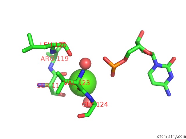
Mono view
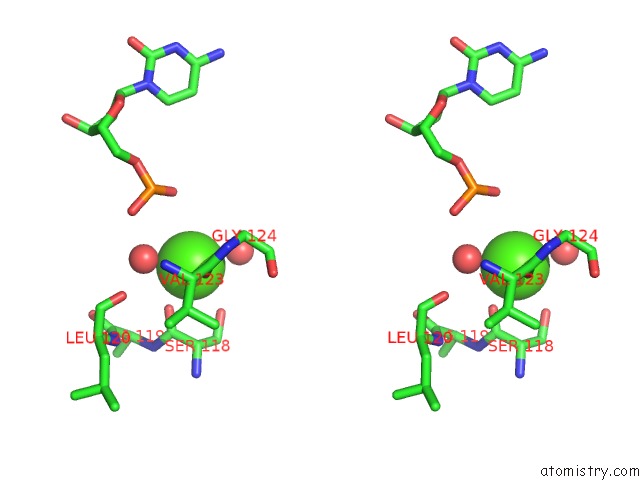
Stereo pair view
Mono view
Stereo pair view
A full contact list of Calcium with other atoms in the Ca binding
site number 1 of Muty Adenine Glycosylase in Complex with Dna and Soaked Adenine Free Base within 5.0Å range:
|
Reference:
J.C.Fromme,
A.Banerjee,
S.J.Huang,
G.L.Verdine.
Structural Basis For Removal of Adenine Mispaired with 8-Oxoguanine By Muty Adenine Dna Glycosylase Nature V. 427 652 2004.
ISSN: ISSN 0028-0836
PubMed: 14961129
DOI: 10.1038/NATURE02306
Page generated: Fri Jul 12 06:56:07 2024
ISSN: ISSN 0028-0836
PubMed: 14961129
DOI: 10.1038/NATURE02306
Last articles
Zn in 9J0NZn in 9J0O
Zn in 9J0P
Zn in 9FJX
Zn in 9EKB
Zn in 9C0F
Zn in 9CAH
Zn in 9CH0
Zn in 9CH3
Zn in 9CH1