Calcium »
PDB 1vfo-1w7c »
1w20 »
Calcium in PDB 1w20: Structure of Neuraminidase From English Duck Subtype N6 Complexed with 30 Mm Sialic Acid (Nana, NEU5AC), Crystal Soaked For 3 Hours at 291 K
Enzymatic activity of Structure of Neuraminidase From English Duck Subtype N6 Complexed with 30 Mm Sialic Acid (Nana, NEU5AC), Crystal Soaked For 3 Hours at 291 K
All present enzymatic activity of Structure of Neuraminidase From English Duck Subtype N6 Complexed with 30 Mm Sialic Acid (Nana, NEU5AC), Crystal Soaked For 3 Hours at 291 K:
3.2.1.18;
3.2.1.18;
Protein crystallography data
The structure of Structure of Neuraminidase From English Duck Subtype N6 Complexed with 30 Mm Sialic Acid (Nana, NEU5AC), Crystal Soaked For 3 Hours at 291 K, PDB code: 1w20
was solved by
E.Rudino-Pinera,
P.Tunnah,
S.J.Crennell,
R.G.Webster,
W.G.Laver,
E.F.Garman,
with X-Ray Crystallography technique. A brief refinement statistics is given in the table below:
| Resolution Low / High (Å) | 105.41 / 2.08 |
| Space group | P 1 21 1 |
| Cell size a, b, c (Å), α, β, γ (°) | 106.478, 73.995, 106.465, 90.00, 90.50, 90.00 |
| R / Rfree (%) | 15 / 19.5 |
Calcium Binding Sites:
The binding sites of Calcium atom in the Structure of Neuraminidase From English Duck Subtype N6 Complexed with 30 Mm Sialic Acid (Nana, NEU5AC), Crystal Soaked For 3 Hours at 291 K
(pdb code 1w20). This binding sites where shown within
5.0 Angstroms radius around Calcium atom.
In total 4 binding sites of Calcium where determined in the Structure of Neuraminidase From English Duck Subtype N6 Complexed with 30 Mm Sialic Acid (Nana, NEU5AC), Crystal Soaked For 3 Hours at 291 K, PDB code: 1w20:
Jump to Calcium binding site number: 1; 2; 3; 4;
In total 4 binding sites of Calcium where determined in the Structure of Neuraminidase From English Duck Subtype N6 Complexed with 30 Mm Sialic Acid (Nana, NEU5AC), Crystal Soaked For 3 Hours at 291 K, PDB code: 1w20:
Jump to Calcium binding site number: 1; 2; 3; 4;
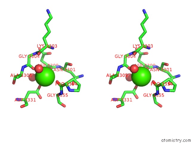
Calcium binding site 1 out of 4 in 1w20
Go back to
Calcium binding site 1 out
of 4 in the Structure of Neuraminidase From English Duck Subtype N6 Complexed with 30 Mm Sialic Acid (Nana, NEU5AC), Crystal Soaked For 3 Hours at 291 K
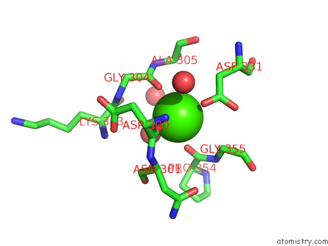
Mono view

Stereo pair view
Mono view
Stereo pair view
A full contact list of Calcium with other atoms in the Ca binding
site number 1 of Structure of Neuraminidase From English Duck Subtype N6 Complexed with 30 Mm Sialic Acid (Nana, NEU5AC), Crystal Soaked For 3 Hours at 291 K within 5.0Å range:
|
Calcium binding site 2 out of 4 in 1w20
Go back to
Calcium binding site 2 out
of 4 in the Structure of Neuraminidase From English Duck Subtype N6 Complexed with 30 Mm Sialic Acid (Nana, NEU5AC), Crystal Soaked For 3 Hours at 291 K
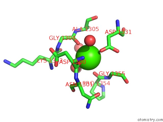
Mono view
Stereo pair view
Mono view
Stereo pair view
A full contact list of Calcium with other atoms in the Ca binding
site number 2 of Structure of Neuraminidase From English Duck Subtype N6 Complexed with 30 Mm Sialic Acid (Nana, NEU5AC), Crystal Soaked For 3 Hours at 291 K within 5.0Å range:
|
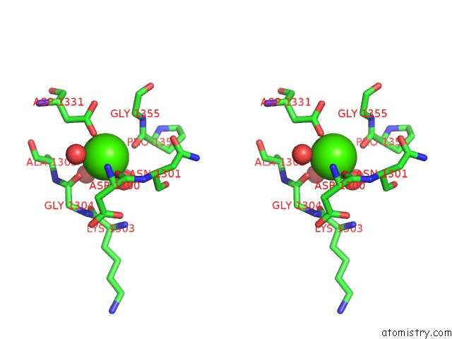
Calcium binding site 3 out of 4 in 1w20
Go back to
Calcium binding site 3 out
of 4 in the Structure of Neuraminidase From English Duck Subtype N6 Complexed with 30 Mm Sialic Acid (Nana, NEU5AC), Crystal Soaked For 3 Hours at 291 K

Mono view

Stereo pair view
Mono view
Stereo pair view
A full contact list of Calcium with other atoms in the Ca binding
site number 3 of Structure of Neuraminidase From English Duck Subtype N6 Complexed with 30 Mm Sialic Acid (Nana, NEU5AC), Crystal Soaked For 3 Hours at 291 K within 5.0Å range:
|
Calcium binding site 4 out of 4 in 1w20
Go back to
Calcium binding site 4 out
of 4 in the Structure of Neuraminidase From English Duck Subtype N6 Complexed with 30 Mm Sialic Acid (Nana, NEU5AC), Crystal Soaked For 3 Hours at 291 K
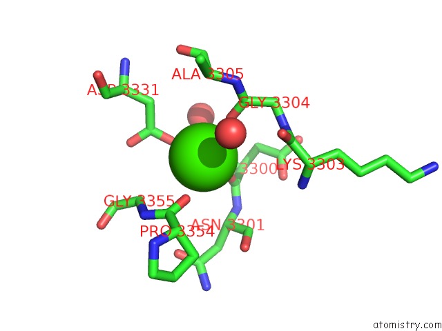
Mono view
Stereo pair view
Mono view
Stereo pair view
A full contact list of Calcium with other atoms in the Ca binding
site number 4 of Structure of Neuraminidase From English Duck Subtype N6 Complexed with 30 Mm Sialic Acid (Nana, NEU5AC), Crystal Soaked For 3 Hours at 291 K within 5.0Å range:
|
Reference:
E.Rudino-Pinera,
P.Tunnah,
S.J.Crennell,
R.G.Webster,
W.G.Laver,
E.F.Garman.
The Crystal Structure of Type A Influenza Virus Neuraminidase of the N6 Subtype Reveals the Existence of Two Separate NEU5AC Binding Sites To Be Published.
Page generated: Fri Jul 12 06:58:41 2024
Last articles
Zn in 9JYWZn in 9IR4
Zn in 9IR3
Zn in 9GMX
Zn in 9GMW
Zn in 9JEJ
Zn in 9ERF
Zn in 9ERE
Zn in 9EGV
Zn in 9EGW