Atomistry » Calcium » PDB 1vfo-1w7c » 1w32
Atomistry »
  Calcium »
    PDB 1vfo-1w7c »
      1w32 »

Calcium in PDB 1w32: The 3-Dimensional Structure of A Thermostable Mutant of A Xylanase (XYN10A) From Cellvibrio Japonicus

Enzymatic activity of The 3-Dimensional Structure of A Thermostable Mutant of A Xylanase (XYN10A) From Cellvibrio Japonicus

All present enzymatic activity of The 3-Dimensional Structure of A Thermostable Mutant of A Xylanase (XYN10A) From Cellvibrio Japonicus:
3.2.1.8;

Protein crystallography data

The structure of The 3-Dimensional Structure of A Thermostable Mutant of A Xylanase (XYN10A) From Cellvibrio Japonicus, PDB code: 1w32 was solved by S.Andrews, E.J.Taylor, G.N.Pell, F.Vincent, V.M.A.Ducros, G.J.Davies, J.H.Lakey, H.J.Gilbert, with X-Ray Crystallography technique. A brief refinement statistics is given in the table below:

Resolution Low / High (Å) 81.65 / 1.2
Space group P 43 21 2
Cell size a, b, c (Å), α, β, γ (°) 95.343, 95.343, 150.273, 90.00, 90.00, 90.00
R / Rfree (%) 12.1 / 14.4

Calcium Binding Sites:

The binding sites of Calcium atom in the The 3-Dimensional Structure of A Thermostable Mutant of A Xylanase (XYN10A) From Cellvibrio Japonicus (pdb code 1w32). This binding sites where shown within 5.0 Angstroms radius around Calcium atom.
In total 3 binding sites of Calcium where determined in the The 3-Dimensional Structure of A Thermostable Mutant of A Xylanase (XYN10A) From Cellvibrio Japonicus, PDB code: 1w32:
Jump to Calcium binding site number: 1; 2; 3;

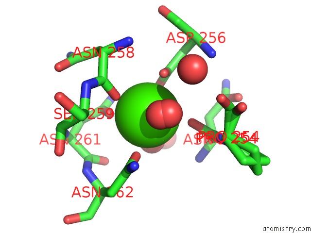
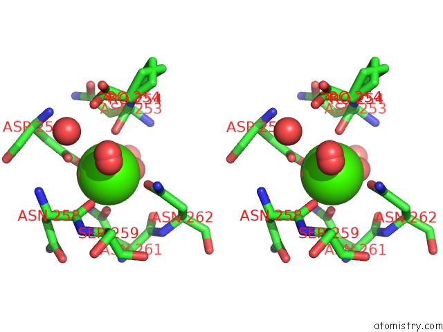
Calcium binding site 1 out of 3 in 1w32

Go back to Calcium Binding Sites List in 1w32
Calcium binding site 1 out of 3 in the The 3-Dimensional Structure of A Thermostable Mutant of A Xylanase (XYN10A) From Cellvibrio Japonicus


Mono view


Stereo pair view

A full contact list of Calcium with other atoms in the Ca binding site number 1 of The 3-Dimensional Structure of A Thermostable Mutant of A Xylanase (XYN10A) From Cellvibrio Japonicus within 5.0Å range:
probe atom residue distance (Å) B Occ
A:Ca1347

b:8.9
occ:0.50
CA A:CA1347 0.0 8.9 0.5
CA A:CA1347 0.6 10.0 0.5
OD1 A:ASP256 1.9 13.3 0.5
OD2 A:ASP256 2.2 12.0 0.5
O A:ASN258 2.3 11.5 0.5
CG A:ASP256 2.3 13.2 0.5
OD1 A:ASN261 2.4 9.3 0.5
OD2 A:ASP256 2.4 11.1 0.5
O A:ASN258 2.4 10.5 0.5
O A:ASN253 2.4 7.0 0.5
ND2 A:ASN262 2.4 5.5 0.5
O A:HOH2405 2.5 12.2 1.0
OD1 A:ASP256 2.6 10.3 0.5
OD1 A:ASN261 2.6 12.7 0.5
O A:HOH2385 2.7 12.2 1.0
O A:ASN253 2.8 12.0 0.5
CG A:ASP256 2.8 11.1 0.5
ND2 A:ASN262 3.0 9.8 0.5
C A:ASN258 3.4 11.0 0.5
CG A:ASN261 3.4 10.1 0.5
CG A:ASN262 3.5 8.3 0.5
CG A:ASN261 3.5 13.5 0.5
C A:ASN258 3.6 11.1 0.5
C A:ASN253 3.6 7.2 0.5
ND2 A:ASN261 3.8 14.6 0.5
OD1 A:ASN262 3.8 10.5 0.5
ND2 A:ASN261 3.8 9.9 0.5
CB A:ASP256 3.8 13.8 0.5
C A:ASN253 4.0 11.9 0.5
CG A:ASN262 4.0 11.0 0.5
CA A:ASN258 4.0 11.1 0.5
N A:ASN258 4.1 11.9 0.5
CB A:ASN258 4.1 11.5 0.5
CB A:ASN258 4.3 11.4 0.5
CA A:ASN258 4.3 11.2 0.5
CB A:ASP256 4.3 11.4 0.5
OD1 A:ASN262 4.3 11.2 0.5
N A:ASN258 4.4 12.3 0.5
CA A:ASN253 4.4 7.0 0.5
CB A:ASN253 4.4 7.0 0.5
N A:ASP256 4.4 14.3 0.5
C A:ASN261 4.4 9.3 0.5
O A:ASN261 4.4 10.1 0.5
N A:ASN253 4.4 6.5 0.5
N A:SER259 4.5 11.2 0.5
CA A:ASP256 4.6 13.9 0.5
N A:SER259 4.6 11.2 0.5
N A:PRO254 4.6 8.0 0.5
N A:ASN262 4.6 8.7 0.5
O A:HOH2391 4.7 14.1 0.5
CA A:SER259 4.7 11.6 0.5
CB A:ASN261 4.7 10.2 0.5
CA A:PRO254 4.7 8.8 0.5
CB A:ASN253 4.7 11.6 0.5
CA A:SER259 4.7 12.0 0.5
CA A:ASN253 4.7 11.4 0.5
CB A:ASN262 4.8 8.1 0.5
N A:ASN253 4.8 10.6 0.5
C A:PRO254 4.8 9.2 0.5
N A:ASP256 4.8 12.2 0.5
C A:ASP256 4.8 14.0 0.5
N A:ASN261 4.8 10.9 0.5
CG A:ASN258 4.9 12.0 0.5
CA A:ASN261 4.9 10.1 0.5
O A:HOH2390 4.9 17.2 0.5
CA A:ASN262 4.9 8.1 0.5
CB A:ASN261 4.9 12.7 0.5
N A:PRO254 5.0 12.9 0.5
O A:HOH2392 5.0 26.5 1.0

Calcium binding site 2 out of 3 in 1w32

Go back to Calcium Binding Sites List in 1w32
Calcium binding site 2 out of 3 in the The 3-Dimensional Structure of A Thermostable Mutant of A Xylanase (XYN10A) From Cellvibrio Japonicus


Mono view


Stereo pair view

A full contact list of Calcium with other atoms in the Ca binding site number 2 of The 3-Dimensional Structure of A Thermostable Mutant of A Xylanase (XYN10A) From Cellvibrio Japonicus within 5.0Å range:
probe atom residue distance (Å) B Occ
A:Ca1347

b:10.0
occ:0.50
CA A:CA1347 0.0 10.0 0.5
CA A:CA1347 0.6 8.9 0.5
ND2 A:ASN262 1.9 5.5 0.5
O A:HOH2385 2.3 12.2 1.0
ND2 A:ASN262 2.4 9.8 0.5
O A:ASN253 2.4 7.0 0.5
OD1 A:ASN261 2.4 9.3 0.5
O A:ASN258 2.4 11.5 0.5
OD1 A:ASN261 2.4 12.7 0.5
OD1 A:ASP256 2.5 13.3 0.5
OD2 A:ASP256 2.6 12.0 0.5
O A:HOH2405 2.6 12.2 1.0
O A:ASN258 2.6 10.5 0.5
O A:ASN253 2.7 12.0 0.5
CG A:ASP256 2.8 13.2 0.5
OD2 A:ASP256 2.9 11.1 0.5
CG A:ASN262 3.0 8.3 0.5
OD1 A:ASP256 3.0 10.3 0.5
CG A:ASP256 3.3 11.1 0.5
OD1 A:ASN262 3.5 10.5 0.5
CG A:ASN261 3.5 13.5 0.5
CG A:ASN261 3.5 10.1 0.5
C A:ASN253 3.5 7.2 0.5
CG A:ASN262 3.5 11.0 0.5
C A:ASN258 3.6 11.0 0.5
ND2 A:ASN261 3.8 14.6 0.5
C A:ASN253 3.8 11.9 0.5
C A:ASN258 3.9 11.1 0.5
OD1 A:ASN262 3.9 11.2 0.5
ND2 A:ASN261 4.0 9.9 0.5
O A:ASN261 4.1 10.1 0.5
C A:ASN261 4.1 9.3 0.5
N A:ASN253 4.1 6.5 0.5
N A:ASN262 4.2 8.7 0.5
CB A:ASN262 4.2 8.1 0.5
CA A:ASN253 4.2 7.0 0.5
CB A:ASP256 4.3 13.8 0.5
CA A:ASN262 4.4 8.1 0.5
CB A:ASN253 4.4 7.0 0.5
CA A:ASN258 4.4 11.1 0.5
N A:ASN253 4.5 10.6 0.5
N A:PRO254 4.5 8.0 0.5
CB A:ASN258 4.5 11.5 0.5
N A:ASN258 4.6 11.9 0.5
CA A:ASN253 4.6 11.4 0.5
N A:ASN262 4.6 11.6 0.5
N A:SER259 4.6 11.2 0.5
CA A:PRO254 4.6 8.8 0.5
O A:HOH2391 4.6 14.1 0.5
CA A:SER259 4.6 11.6 0.5
CB A:ASN261 4.7 10.2 0.5
CB A:ASN253 4.7 11.6 0.5
CA A:ASN261 4.7 10.1 0.5
CB A:ASN258 4.7 11.4 0.5
N A:ASN261 4.7 10.9 0.5
CA A:ASN258 4.7 11.2 0.5
CA A:SER259 4.7 12.0 0.5
C A:ASN261 4.8 12.7 0.5
CB A:ASN262 4.8 10.7 0.5
N A:SER259 4.8 11.2 0.5
CB A:ASP256 4.8 11.4 0.5
CB A:ASN261 4.8 12.7 0.5
N A:PRO254 4.8 12.9 0.5
N A:ASP256 4.8 14.3 0.5
N A:ASN258 4.9 12.3 0.5
CA A:ASN262 4.9 10.7 0.5
O A:HOH2383 4.9 19.6 0.5
C A:PRO254 4.9 9.2 0.5
CA A:PRO254 5.0 13.6 0.5

Calcium binding site 3 out of 3 in 1w32

Go back to Calcium Binding Sites List in 1w32
Calcium binding site 3 out of 3 in the The 3-Dimensional Structure of A Thermostable Mutant of A Xylanase (XYN10A) From Cellvibrio Japonicus


Mono view


Stereo pair view

A full contact list of Calcium with other atoms in the Ca binding site number 3 of The 3-Dimensional Structure of A Thermostable Mutant of A Xylanase (XYN10A) From Cellvibrio Japonicus within 5.0Å range:
probe atom residue distance (Å) B Occ
B:Ca1347

b:6.8
occ:1.00
OD1 B:ASN262 2.4 7.7 1.0
OD1 B:ASN261 2.4 8.8 1.0
O B:ASN258 2.4 8.1 1.0
OD1 B:ASP256 2.5 7.7 1.0
O B:ASN253 2.5 6.8 1.0
O B:HOH2389 2.5 4.6 0.5
O B:HOH2398 2.5 7.9 1.0
OD2 B:ASP256 2.6 8.6 1.0
O B:HOH2381 2.7 8.7 0.5
CG B:ASP256 2.9 8.3 1.0
CG B:ASN261 3.4 9.3 1.0
CG B:ASN262 3.4 7.1 1.0
C B:ASN258 3.6 7.1 1.0
C B:ASN253 3.6 6.8 1.0
ND2 B:ASN261 3.8 12.7 1.0
ND2 B:ASN262 3.9 7.6 1.0
CB B:ASP256 4.3 8.6 1.0
O B:ASN261 4.3 11.2 1.0
CA B:ASN258 4.4 7.8 1.0
CB B:ASN258 4.4 8.8 1.0
C B:ASN261 4.4 9.2 1.0
CA B:ASN253 4.4 6.9 1.0
N B:ASN253 4.4 7.1 1.0
N B:ASN258 4.4 7.7 1.0
CB B:ASN253 4.5 8.0 1.0
N B:ASN262 4.6 8.0 1.0
N B:SER259 4.6 8.0 1.0
N B:PRO254 4.6 6.4 0.5
O B:HOH2397 4.6 10.5 0.5
N B:PRO254 4.6 7.5 0.5
O B:HOH2383 4.7 10.0 0.5
CA B:PRO254 4.7 8.4 0.5
CB B:ASN262 4.7 7.5 1.0
CB B:ASN261 4.7 9.8 1.0
CA B:SER259 4.7 9.5 1.0
CA B:PRO254 4.8 6.9 0.5
N B:ASP256 4.8 8.4 1.0
CA B:ASN262 4.9 7.8 1.0
C B:PRO254 4.9 6.9 0.5
N B:ASN261 4.9 9.4 1.0
CA B:ASN261 4.9 9.8 1.0
O B:HOH2380 5.0 19.1 0.5

Reference:

S.Andrews, E.J.Taylor, G.N.Pell, F.Vincent, V.M.A.Ducros, G.J.Davies, J.H.Lakey, H.J.Gilbert. The Use of Forced Protein Evolution to Investigate and Improve Stability of Family 10 Xylanases: the Production of CA2+-Independent Stable Xylanases J.Biol.Chem. V. 279 54369 2004.
ISSN: ISSN 0021-9258
PubMed: 15452124
DOI: 10.1074/JBC.M409044200
Page generated: Tue Jul 8 03:06:12 2025

Last articles

F in 7NWA
F in 7NVN
F in 7NVM
F in 7NW2
F in 7NW0
F in 7NVL
F in 7NVX
F in 7NVV
F in 7NVO
F in 7NTH
© Copyright 2008-2020 by atomistry.com
Home   |    Site Map   |    Copyright   |    Contact us   |    Privacy