Calcium »
PDB 1vfo-1w7c »
1w3w »
Calcium in PDB 1w3w: The 2.1 Angstroem Resolution Structure of Annexin A8
Protein crystallography data
The structure of The 2.1 Angstroem Resolution Structure of Annexin A8, PDB code: 1w3w
was solved by
S.Rety,
J.Sopkova-De Oliveira Santos,
M.Renouard,
A.Lewit-Bentley,
with X-Ray Crystallography technique. A brief refinement statistics is given in the table below:
| Resolution Low / High (Å) | 23.31 / 1.99 |
| Space group | P 1 21 1 |
| Cell size a, b, c (Å), α, β, γ (°) | 50.761, 65.581, 59.229, 90.00, 100.70, 90.00 |
| R / Rfree (%) | 17.5 / 21.8 |
Calcium Binding Sites:
The binding sites of Calcium atom in the The 2.1 Angstroem Resolution Structure of Annexin A8
(pdb code 1w3w). This binding sites where shown within
5.0 Angstroms radius around Calcium atom.
In total only one binding site of Calcium was determined in the The 2.1 Angstroem Resolution Structure of Annexin A8, PDB code: 1w3w:
In total only one binding site of Calcium was determined in the The 2.1 Angstroem Resolution Structure of Annexin A8, PDB code: 1w3w:
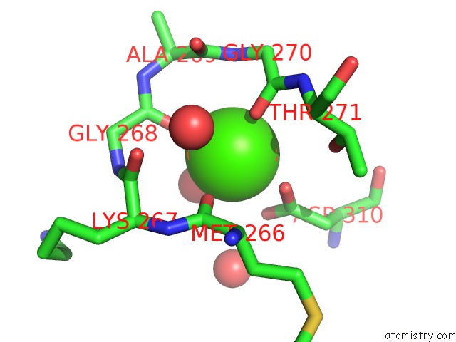
Calcium binding site 1 out of 1 in 1w3w
Go back to
Calcium binding site 1 out
of 1 in the The 2.1 Angstroem Resolution Structure of Annexin A8
Mono view

Stereo pair view
Mono view
Stereo pair view
A full contact list of Calcium with other atoms in the Ca binding
site number 1 of The 2.1 Angstroem Resolution Structure of Annexin A8 within 5.0Å range:
|
Reference:
S.Rety,
J.Sopkova-De Oliveira Santos,
L.Dreyfuss,
K.Blondeau,
K.Hofbauerova,
C.Raguenes-Nicol,
D.Kerboeuf,
M.Renouard,
F.Russo-Marie,
A.Lewit-Bentley.
The Crystal Structure of Annexin A8 Is Similar to That of Annexin A3 J.Mol.Biol. V. 345 1131 2005.
ISSN: ISSN 0022-2836
PubMed: 15644210
DOI: 10.1016/J.JMB.2004.11.015
Page generated: Tue Jul 8 03:06:56 2025
ISSN: ISSN 0022-2836
PubMed: 15644210
DOI: 10.1016/J.JMB.2004.11.015
Last articles
Ca in 3AK5Ca in 3AJ9
Ca in 3AIE
Ca in 3AJ8
Ca in 3AIC
Ca in 3AJ7
Ca in 3AIB
Ca in 3AII
Ca in 3AIG
Ca in 3AI7