Calcium »
PDB 1vfo-1w7c »
1w52 »
Calcium in PDB 1w52: Crystal Structure of A Proteolyzed Form of Pancreatic Lipase Related Protein 2 From Horse
Protein crystallography data
The structure of Crystal Structure of A Proteolyzed Form of Pancreatic Lipase Related Protein 2 From Horse, PDB code: 1w52
was solved by
J.M.Mancheno,
S.Jayne,
B.Kerfelec,
C.Chapus,
I.Crenon,
J.A.Hermoso,
with X-Ray Crystallography technique. A brief refinement statistics is given in the table below:
| Resolution Low / High (Å) | 26.17 / 2.99 |
| Space group | P 32 2 1 |
| Cell size a, b, c (Å), α, β, γ (°) | 128.426, 128.426, 85.818, 90.00, 90.00, 120.00 |
| R / Rfree (%) | 23.1 / 29.4 |
Calcium Binding Sites:
The binding sites of Calcium atom in the Crystal Structure of A Proteolyzed Form of Pancreatic Lipase Related Protein 2 From Horse
(pdb code 1w52). This binding sites where shown within
5.0 Angstroms radius around Calcium atom.
In total 2 binding sites of Calcium where determined in the Crystal Structure of A Proteolyzed Form of Pancreatic Lipase Related Protein 2 From Horse, PDB code: 1w52:
Jump to Calcium binding site number: 1; 2;
In total 2 binding sites of Calcium where determined in the Crystal Structure of A Proteolyzed Form of Pancreatic Lipase Related Protein 2 From Horse, PDB code: 1w52:
Jump to Calcium binding site number: 1; 2;
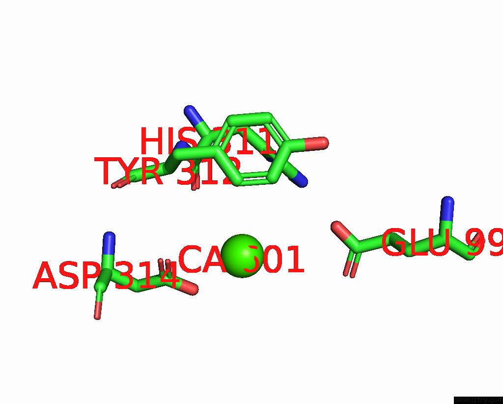
Calcium binding site 1 out of 2 in 1w52
Go back to
Calcium binding site 1 out
of 2 in the Crystal Structure of A Proteolyzed Form of Pancreatic Lipase Related Protein 2 From Horse
Mono view
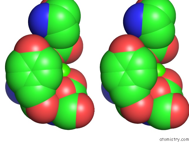
Stereo pair view
Mono view
Stereo pair view
A full contact list of Calcium with other atoms in the Ca binding
site number 1 of Crystal Structure of A Proteolyzed Form of Pancreatic Lipase Related Protein 2 From Horse within 5.0Å range:
|
Calcium binding site 2 out of 2 in 1w52
Go back to
Calcium binding site 2 out
of 2 in the Crystal Structure of A Proteolyzed Form of Pancreatic Lipase Related Protein 2 From Horse
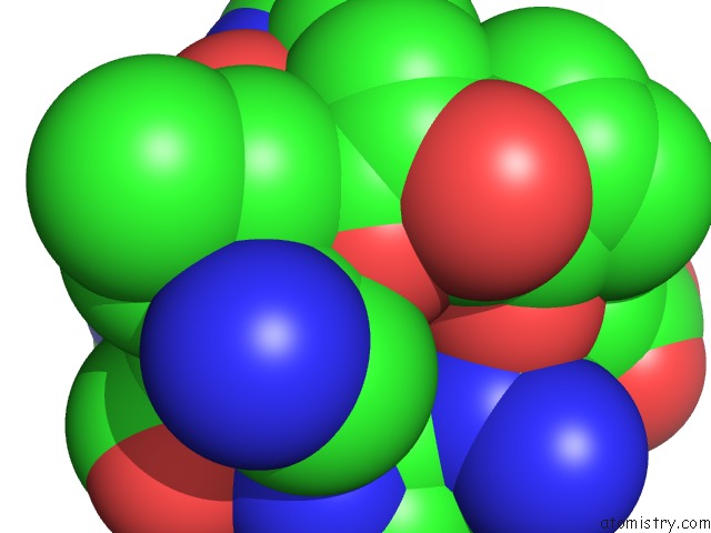
Mono view
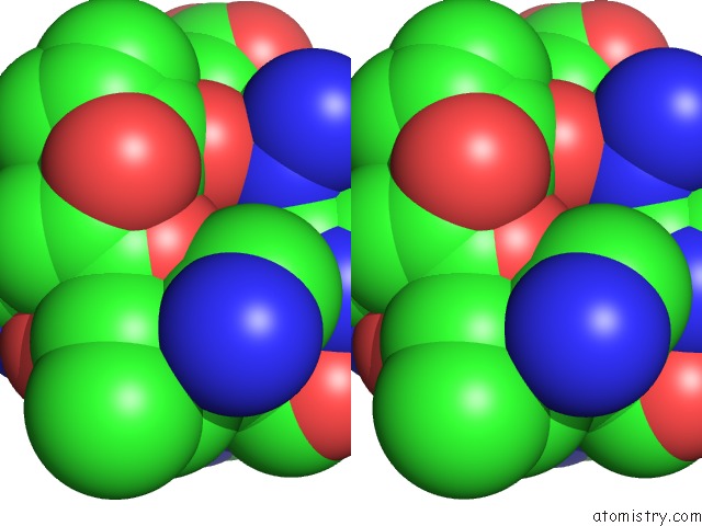
Stereo pair view
Mono view
Stereo pair view
A full contact list of Calcium with other atoms in the Ca binding
site number 2 of Crystal Structure of A Proteolyzed Form of Pancreatic Lipase Related Protein 2 From Horse within 5.0Å range:
|
Reference:
J.M.Mancheno,
S.Jayne,
B.Kerfelec,
C.Chapus,
I.Crenon,
J.A.Hermoso.
Crystalization of A Proteolyzed Form of the Horse Pancreatic Lipase-Related Protein 2: Structural Basis For the Specific Detergent Requirement. Acta Crystallogr.,Sect.D V. 60 2107 2004.
ISSN: ISSN 0907-4449
PubMed: 15502342
DOI: 10.1107/S0907444904024229
Page generated: Fri Jul 12 07:04:25 2024
ISSN: ISSN 0907-4449
PubMed: 15502342
DOI: 10.1107/S0907444904024229
Last articles
Zn in 9JYWZn in 9IR4
Zn in 9IR3
Zn in 9GMX
Zn in 9GMW
Zn in 9JEJ
Zn in 9ERF
Zn in 9ERE
Zn in 9EGV
Zn in 9EGW