Calcium »
PDB 1w7x-1wws »
1wme »
Calcium in PDB 1wme: Crystal Structure of Alkaline Serine Protease Kp-43 From Bacillus Sp. Ksm-KP43 (1.50 Angstrom, 293 K)
Protein crystallography data
The structure of Crystal Structure of Alkaline Serine Protease Kp-43 From Bacillus Sp. Ksm-KP43 (1.50 Angstrom, 293 K), PDB code: 1wme
was solved by
T.Nonaka,
M.Fujihashi,
A.Kita,
K.Saeki,
S.Ito,
K.Horikoshi,
K.Miki,
with X-Ray Crystallography technique. A brief refinement statistics is given in the table below:
| Resolution Low / High (Å) | 26.00 / 1.50 |
| Space group | C 2 2 21 |
| Cell size a, b, c (Å), α, β, γ (°) | 43.539, 110.386, 168.960, 90.00, 90.00, 90.00 |
| R / Rfree (%) | 12.6 / 16.2 |
Calcium Binding Sites:
The binding sites of Calcium atom in the Crystal Structure of Alkaline Serine Protease Kp-43 From Bacillus Sp. Ksm-KP43 (1.50 Angstrom, 293 K)
(pdb code 1wme). This binding sites where shown within
5.0 Angstroms radius around Calcium atom.
In total 3 binding sites of Calcium where determined in the Crystal Structure of Alkaline Serine Protease Kp-43 From Bacillus Sp. Ksm-KP43 (1.50 Angstrom, 293 K), PDB code: 1wme:
Jump to Calcium binding site number: 1; 2; 3;
In total 3 binding sites of Calcium where determined in the Crystal Structure of Alkaline Serine Protease Kp-43 From Bacillus Sp. Ksm-KP43 (1.50 Angstrom, 293 K), PDB code: 1wme:
Jump to Calcium binding site number: 1; 2; 3;
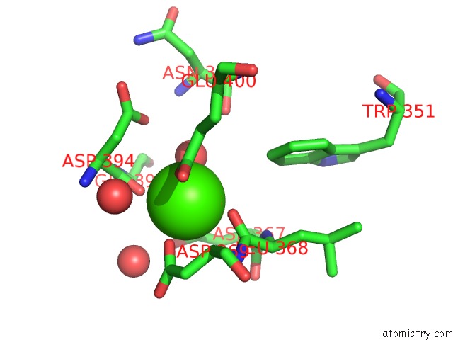
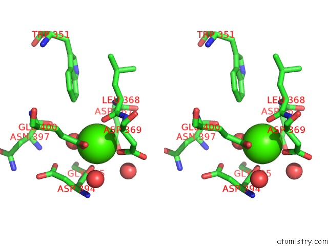
Calcium binding site 1 out of 3 in 1wme
Go back to
Calcium binding site 1 out
of 3 in the Crystal Structure of Alkaline Serine Protease Kp-43 From Bacillus Sp. Ksm-KP43 (1.50 Angstrom, 293 K)
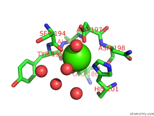
Mono view
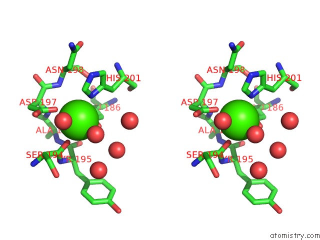
Stereo pair view
Mono view
Stereo pair view
A full contact list of Calcium with other atoms in the Ca binding
site number 1 of Crystal Structure of Alkaline Serine Protease Kp-43 From Bacillus Sp. Ksm-KP43 (1.50 Angstrom, 293 K) within 5.0Å range:
|
Calcium binding site 2 out of 3 in 1wme
Go back to
Calcium binding site 2 out
of 3 in the Crystal Structure of Alkaline Serine Protease Kp-43 From Bacillus Sp. Ksm-KP43 (1.50 Angstrom, 293 K)
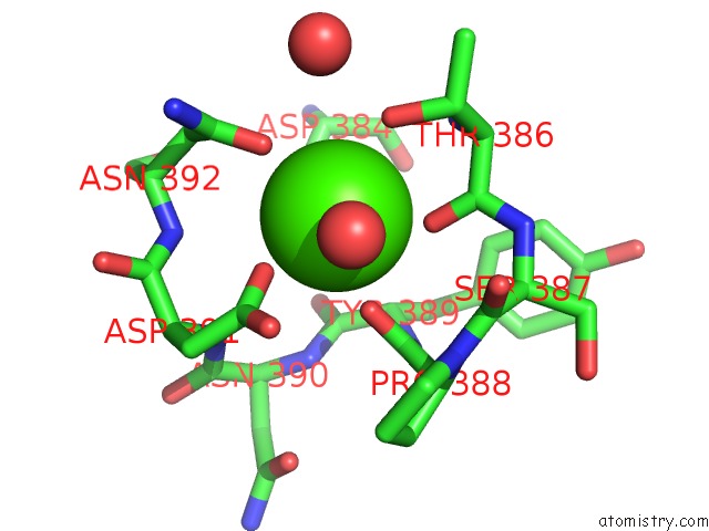
Mono view
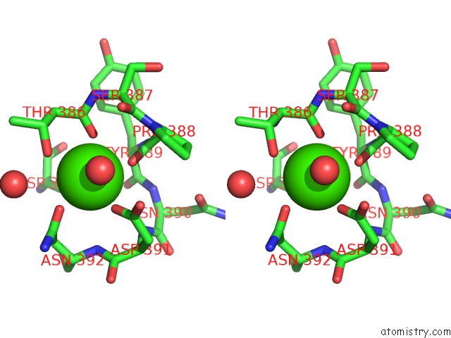
Stereo pair view
Mono view
Stereo pair view
A full contact list of Calcium with other atoms in the Ca binding
site number 2 of Crystal Structure of Alkaline Serine Protease Kp-43 From Bacillus Sp. Ksm-KP43 (1.50 Angstrom, 293 K) within 5.0Å range:
|
Calcium binding site 3 out of 3 in 1wme
Go back to
Calcium binding site 3 out
of 3 in the Crystal Structure of Alkaline Serine Protease Kp-43 From Bacillus Sp. Ksm-KP43 (1.50 Angstrom, 293 K)
Mono view
Stereo pair view
Mono view
Stereo pair view
A full contact list of Calcium with other atoms in the Ca binding
site number 3 of Crystal Structure of Alkaline Serine Protease Kp-43 From Bacillus Sp. Ksm-KP43 (1.50 Angstrom, 293 K) within 5.0Å range:
|
Reference:
T.Nonaka,
M.Fujihashi,
A.Kita,
K.Saeki,
S.Ito,
K.Horikoshi,
K.Miki.
The Crystal Structure of An Oxidatively Stable Subtilisin-Like Alkaline Serine Protease, Kp-43, with A C-Terminal {Beta}-Barrel Domain J.Biol.Chem. V. 279 47344 2004.
ISSN: ISSN 0021-9258
PubMed: 15342641
DOI: 10.1074/JBC.M409089200
Page generated: Fri Jul 12 07:12:23 2024
ISSN: ISSN 0021-9258
PubMed: 15342641
DOI: 10.1074/JBC.M409089200
Last articles
Zn in 9J0NZn in 9J0O
Zn in 9J0P
Zn in 9FJX
Zn in 9EKB
Zn in 9C0F
Zn in 9CAH
Zn in 9CH0
Zn in 9CH3
Zn in 9CH1