Calcium »
PDB 1w7x-1wws »
1wra »
Calcium in PDB 1wra: Crystal Structure of Phosphorylcholine Esterase Domain of the Virulence Factor Choline Binding Protein E From Streptococcus Pneumoniae
Enzymatic activity of Crystal Structure of Phosphorylcholine Esterase Domain of the Virulence Factor Choline Binding Protein E From Streptococcus Pneumoniae
All present enzymatic activity of Crystal Structure of Phosphorylcholine Esterase Domain of the Virulence Factor Choline Binding Protein E From Streptococcus Pneumoniae:
3.1.4.38;
3.1.4.38;
Protein crystallography data
The structure of Crystal Structure of Phosphorylcholine Esterase Domain of the Virulence Factor Choline Binding Protein E From Streptococcus Pneumoniae, PDB code: 1wra
was solved by
G.Garau,
O.Dideberg,
with X-Ray Crystallography technique. A brief refinement statistics is given in the table below:
| Resolution Low / High (Å) | 23.84 / 2.00 |
| Space group | P 41 21 2 |
| Cell size a, b, c (Å), α, β, γ (°) | 98.302, 98.302, 172.600, 90.00, 90.00, 90.00 |
| R / Rfree (%) | 17.4 / 20.4 |
Other elements in 1wra:
The structure of Crystal Structure of Phosphorylcholine Esterase Domain of the Virulence Factor Choline Binding Protein E From Streptococcus Pneumoniae also contains other interesting chemical elements:
| Iron | (Fe) | 4 atoms |
Calcium Binding Sites:
The binding sites of Calcium atom in the Crystal Structure of Phosphorylcholine Esterase Domain of the Virulence Factor Choline Binding Protein E From Streptococcus Pneumoniae
(pdb code 1wra). This binding sites where shown within
5.0 Angstroms radius around Calcium atom.
In total 4 binding sites of Calcium where determined in the Crystal Structure of Phosphorylcholine Esterase Domain of the Virulence Factor Choline Binding Protein E From Streptococcus Pneumoniae, PDB code: 1wra:
Jump to Calcium binding site number: 1; 2; 3; 4;
In total 4 binding sites of Calcium where determined in the Crystal Structure of Phosphorylcholine Esterase Domain of the Virulence Factor Choline Binding Protein E From Streptococcus Pneumoniae, PDB code: 1wra:
Jump to Calcium binding site number: 1; 2; 3; 4;
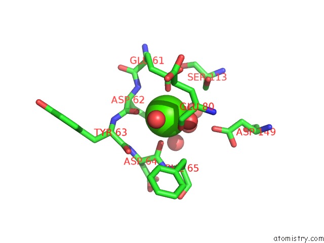
Calcium binding site 1 out of 4 in 1wra
Go back to
Calcium binding site 1 out
of 4 in the Crystal Structure of Phosphorylcholine Esterase Domain of the Virulence Factor Choline Binding Protein E From Streptococcus Pneumoniae
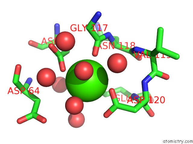
Mono view
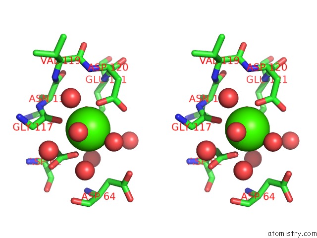
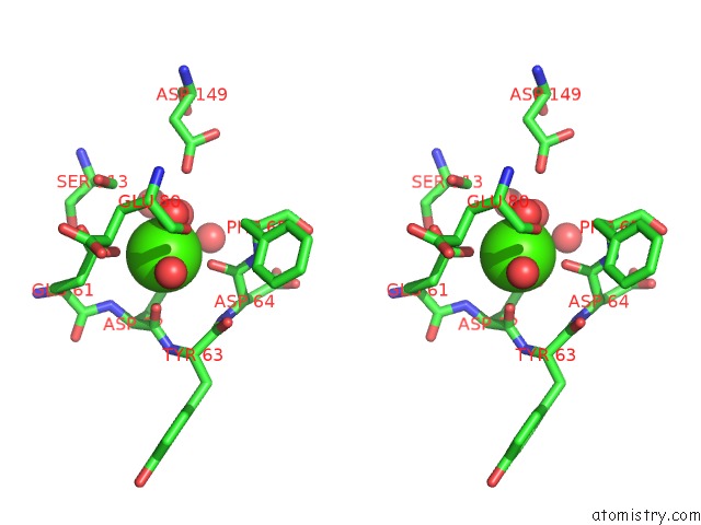
Stereo pair view
Mono view
Stereo pair view
A full contact list of Calcium with other atoms in the Ca binding
site number 1 of Crystal Structure of Phosphorylcholine Esterase Domain of the Virulence Factor Choline Binding Protein E From Streptococcus Pneumoniae within 5.0Å range:
|
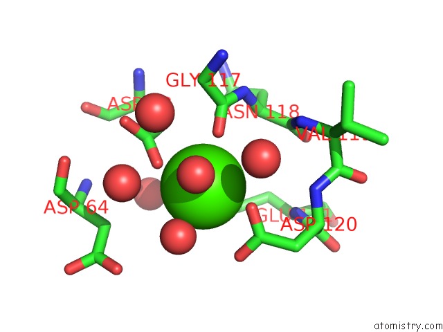
Calcium binding site 2 out of 4 in 1wra
Go back to
Calcium binding site 2 out
of 4 in the Crystal Structure of Phosphorylcholine Esterase Domain of the Virulence Factor Choline Binding Protein E From Streptococcus Pneumoniae
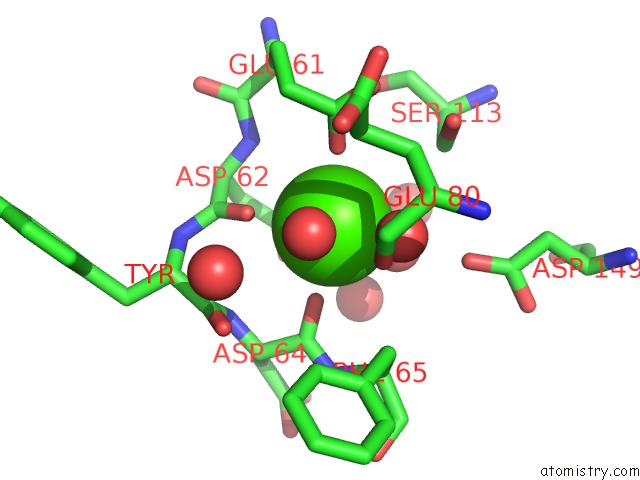
Mono view
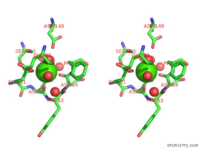
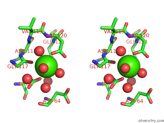
Stereo pair view
Mono view
Stereo pair view
A full contact list of Calcium with other atoms in the Ca binding
site number 2 of Crystal Structure of Phosphorylcholine Esterase Domain of the Virulence Factor Choline Binding Protein E From Streptococcus Pneumoniae within 5.0Å range:
|
Calcium binding site 3 out of 4 in 1wra
Go back to
Calcium binding site 3 out
of 4 in the Crystal Structure of Phosphorylcholine Esterase Domain of the Virulence Factor Choline Binding Protein E From Streptococcus Pneumoniae
Mono view
Stereo pair view
Mono view
Stereo pair view
A full contact list of Calcium with other atoms in the Ca binding
site number 3 of Crystal Structure of Phosphorylcholine Esterase Domain of the Virulence Factor Choline Binding Protein E From Streptococcus Pneumoniae within 5.0Å range:
|
Calcium binding site 4 out of 4 in 1wra
Go back to
Calcium binding site 4 out
of 4 in the Crystal Structure of Phosphorylcholine Esterase Domain of the Virulence Factor Choline Binding Protein E From Streptococcus Pneumoniae
Mono view
Stereo pair view
Mono view
Stereo pair view
A full contact list of Calcium with other atoms in the Ca binding
site number 4 of Crystal Structure of Phosphorylcholine Esterase Domain of the Virulence Factor Choline Binding Protein E From Streptococcus Pneumoniae within 5.0Å range:
|
Reference:
G.Garau,
D.Lemaire,
T.Vernet,
O.Dideberg,
A.M.Di Guilmi.
Crystal Structure of Phosphorylcholine Esterase Domain of the Virulence Factor Choline-Binding Protein E From Streptococcus Pneumoniae: New Structural Features Among the Metallo-Beta-Lactamase Superfamily J.Biol.Chem. V. 280 28591 2005.
ISSN: ISSN 0021-9258
PubMed: 15908436
DOI: 10.1074/JBC.M502744200
Page generated: Tue Jul 8 03:16:13 2025
ISSN: ISSN 0021-9258
PubMed: 15908436
DOI: 10.1074/JBC.M502744200
Last articles
Fe in 2YXOFe in 2YRS
Fe in 2YXC
Fe in 2YNM
Fe in 2YVJ
Fe in 2YP1
Fe in 2YU2
Fe in 2YU1
Fe in 2YQB
Fe in 2YOO