Calcium »
PDB 1wy9-1xjo »
1x1j »
Calcium in PDB 1x1j: Crystal Structure of Xanthan Lyase (N194A) with A Substrate.
Enzymatic activity of Crystal Structure of Xanthan Lyase (N194A) with A Substrate.
All present enzymatic activity of Crystal Structure of Xanthan Lyase (N194A) with A Substrate.:
4.2.2.12;
4.2.2.12;
Protein crystallography data
The structure of Crystal Structure of Xanthan Lyase (N194A) with A Substrate., PDB code: 1x1j
was solved by
Y.Maruyama,
W.Hashimoto,
B.Mikami,
K.Murata,
with X-Ray Crystallography technique. A brief refinement statistics is given in the table below:
| Resolution Low / High (Å) | 45.01 / 2.10 |
| Space group | P 1 21 1 |
| Cell size a, b, c (Å), α, β, γ (°) | 53.747, 90.026, 74.249, 90.00, 91.06, 90.00 |
| R / Rfree (%) | 17.9 / 23.6 |
Calcium Binding Sites:
The binding sites of Calcium atom in the Crystal Structure of Xanthan Lyase (N194A) with A Substrate.
(pdb code 1x1j). This binding sites where shown within
5.0 Angstroms radius around Calcium atom.
In total only one binding site of Calcium was determined in the Crystal Structure of Xanthan Lyase (N194A) with A Substrate., PDB code: 1x1j:
In total only one binding site of Calcium was determined in the Crystal Structure of Xanthan Lyase (N194A) with A Substrate., PDB code: 1x1j:
Calcium binding site 1 out of 1 in 1x1j
Go back to
Calcium binding site 1 out
of 1 in the Crystal Structure of Xanthan Lyase (N194A) with A Substrate.
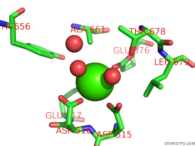
Mono view
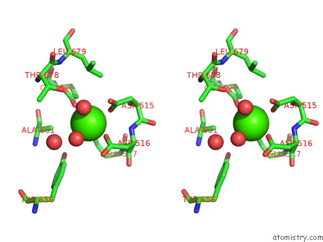
Stereo pair view
Mono view
Stereo pair view
A full contact list of Calcium with other atoms in the Ca binding
site number 1 of Crystal Structure of Xanthan Lyase (N194A) with A Substrate. within 5.0Å range:
|
Reference:
Y.Maruyama,
W.Hashimoto,
B.Mikami,
K.Murata.
Crystal Structure of Bacillus Sp. GL1 Xanthan Lyase Complexed with A Substrate: Insights Into the Enzyme Reaction Mechanism J.Mol.Biol. V. 350 974 2005.
ISSN: ISSN 0022-2836
PubMed: 15979090
DOI: 10.1016/J.JMB.2005.05.055
Page generated: Fri Jul 12 07:25:30 2024
ISSN: ISSN 0022-2836
PubMed: 15979090
DOI: 10.1016/J.JMB.2005.05.055
Last articles
Zn in 9J0NZn in 9J0O
Zn in 9J0P
Zn in 9FJX
Zn in 9EKB
Zn in 9C0F
Zn in 9CAH
Zn in 9CH0
Zn in 9CH3
Zn in 9CH1