Calcium »
PDB 1wy9-1xjo »
1x9d »
Calcium in PDB 1x9d: Crystal Structure of Human Class I Alpha-1,2-Mannosidase in Complex with Thio-Disaccharide Substrate Analogue
Enzymatic activity of Crystal Structure of Human Class I Alpha-1,2-Mannosidase in Complex with Thio-Disaccharide Substrate Analogue
All present enzymatic activity of Crystal Structure of Human Class I Alpha-1,2-Mannosidase in Complex with Thio-Disaccharide Substrate Analogue:
3.2.1.113;
3.2.1.113;
Protein crystallography data
The structure of Crystal Structure of Human Class I Alpha-1,2-Mannosidase in Complex with Thio-Disaccharide Substrate Analogue, PDB code: 1x9d
was solved by
K.Karaveg,
W.Tempel,
Z.J.Liu,
A.Siriwardena,
K.W.Moremen,
B.C.Wang,
with X-Ray Crystallography technique. A brief refinement statistics is given in the table below:
| Resolution Low / High (Å) | 48.80 / 1.41 |
| Space group | P 1 |
| Cell size a, b, c (Å), α, β, γ (°) | 50.687, 53.878, 56.234, 89.49, 63.60, 62.61 |
| R / Rfree (%) | 14.4 / 16.2 |
Calcium Binding Sites:
The binding sites of Calcium atom in the Crystal Structure of Human Class I Alpha-1,2-Mannosidase in Complex with Thio-Disaccharide Substrate Analogue
(pdb code 1x9d). This binding sites where shown within
5.0 Angstroms radius around Calcium atom.
In total only one binding site of Calcium was determined in the Crystal Structure of Human Class I Alpha-1,2-Mannosidase in Complex with Thio-Disaccharide Substrate Analogue, PDB code: 1x9d:
In total only one binding site of Calcium was determined in the Crystal Structure of Human Class I Alpha-1,2-Mannosidase in Complex with Thio-Disaccharide Substrate Analogue, PDB code: 1x9d:
Calcium binding site 1 out of 1 in 1x9d
Go back to
Calcium binding site 1 out
of 1 in the Crystal Structure of Human Class I Alpha-1,2-Mannosidase in Complex with Thio-Disaccharide Substrate Analogue
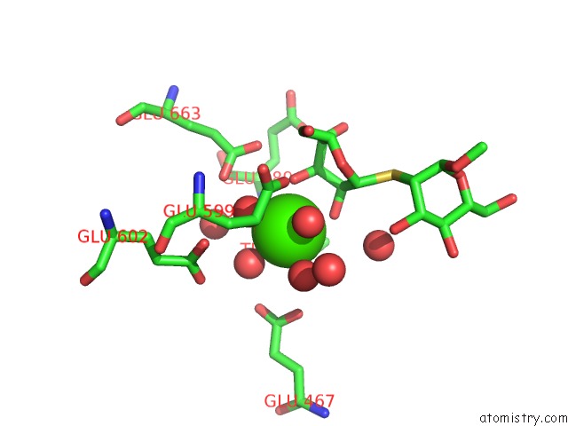
Mono view
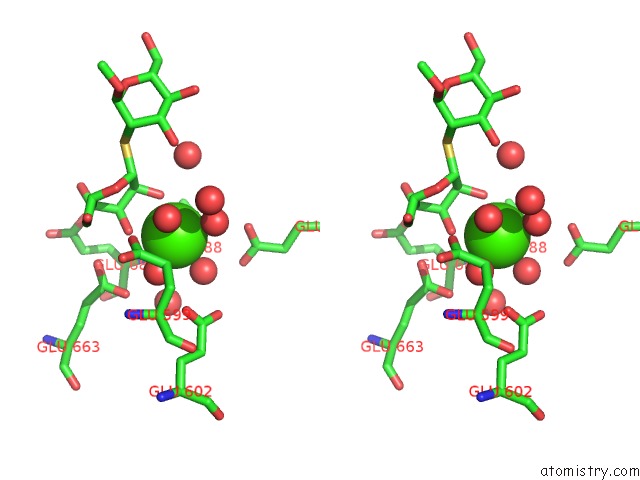
Stereo pair view
Mono view
Stereo pair view
A full contact list of Calcium with other atoms in the Ca binding
site number 1 of Crystal Structure of Human Class I Alpha-1,2-Mannosidase in Complex with Thio-Disaccharide Substrate Analogue within 5.0Å range:
|
Reference:
K.Karaveg,
A.Siriwardena,
W.Tempel,
Z.J.Liu,
J.Glushka,
B.C.Wang,
K.W.Moremen.
Mechanism of Class 1 (Glycosylhydrolase Family 47) {Alpha}-Mannosidases Involved in N-Glycan Processing and Endoplasmic Reticulum Quality Control. J.Biol.Chem. V. 280 16197 2005.
ISSN: ISSN 0021-9258
PubMed: 15713668
DOI: 10.1074/JBC.M500119200
Page generated: Fri Jul 12 07:27:58 2024
ISSN: ISSN 0021-9258
PubMed: 15713668
DOI: 10.1074/JBC.M500119200
Last articles
Zn in 9J0NZn in 9J0O
Zn in 9J0P
Zn in 9FJX
Zn in 9EKB
Zn in 9C0F
Zn in 9CAH
Zn in 9CH0
Zn in 9CH3
Zn in 9CH1