Calcium »
PDB 1wy9-1xjo »
1xhb »
Calcium in PDB 1xhb: The Crystal Structure of Udp-Galnac: Polypeptide Alpha-N- Acetylgalactosaminyltransferase-T1
Enzymatic activity of The Crystal Structure of Udp-Galnac: Polypeptide Alpha-N- Acetylgalactosaminyltransferase-T1
All present enzymatic activity of The Crystal Structure of Udp-Galnac: Polypeptide Alpha-N- Acetylgalactosaminyltransferase-T1:
2.4.1.41;
2.4.1.41;
Protein crystallography data
The structure of The Crystal Structure of Udp-Galnac: Polypeptide Alpha-N- Acetylgalactosaminyltransferase-T1, PDB code: 1xhb
was solved by
T.A.Fritz,
J.H.Hurley,
L.B.Trinh,
J.Shiloach,
L.A.Tabak,
with X-Ray Crystallography technique. A brief refinement statistics is given in the table below:
| Resolution Low / High (Å) | 46.39 / 2.50 |
| Space group | P 43 |
| Cell size a, b, c (Å), α, β, γ (°) | 65.605, 65.605, 125.947, 90.00, 90.00, 90.00 |
| R / Rfree (%) | 21.8 / 25.5 |
Other elements in 1xhb:
The structure of The Crystal Structure of Udp-Galnac: Polypeptide Alpha-N- Acetylgalactosaminyltransferase-T1 also contains other interesting chemical elements:
| Manganese | (Mn) | 1 atom |
Calcium Binding Sites:
The binding sites of Calcium atom in the The Crystal Structure of Udp-Galnac: Polypeptide Alpha-N- Acetylgalactosaminyltransferase-T1
(pdb code 1xhb). This binding sites where shown within
5.0 Angstroms radius around Calcium atom.
In total 2 binding sites of Calcium where determined in the The Crystal Structure of Udp-Galnac: Polypeptide Alpha-N- Acetylgalactosaminyltransferase-T1, PDB code: 1xhb:
Jump to Calcium binding site number: 1; 2;
In total 2 binding sites of Calcium where determined in the The Crystal Structure of Udp-Galnac: Polypeptide Alpha-N- Acetylgalactosaminyltransferase-T1, PDB code: 1xhb:
Jump to Calcium binding site number: 1; 2;
Calcium binding site 1 out of 2 in 1xhb
Go back to
Calcium binding site 1 out
of 2 in the The Crystal Structure of Udp-Galnac: Polypeptide Alpha-N- Acetylgalactosaminyltransferase-T1
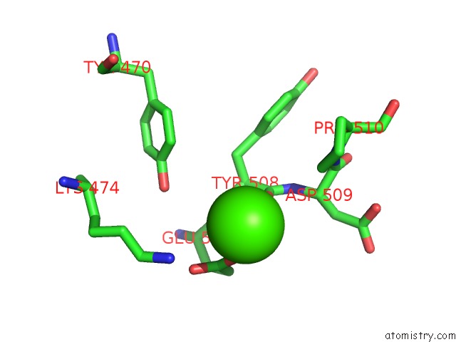
Mono view
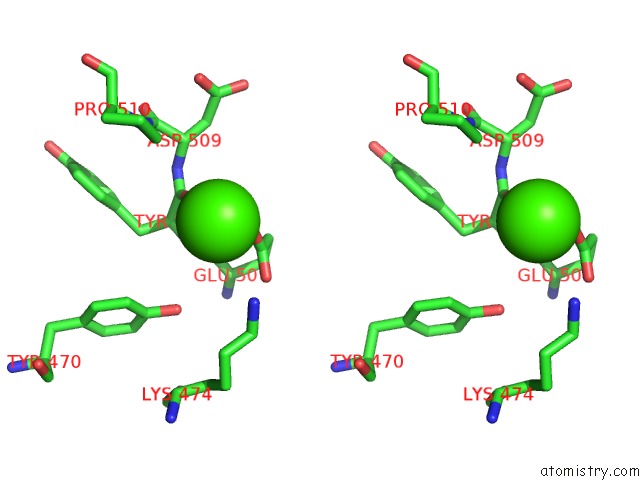
Stereo pair view
Mono view
Stereo pair view
A full contact list of Calcium with other atoms in the Ca binding
site number 1 of The Crystal Structure of Udp-Galnac: Polypeptide Alpha-N- Acetylgalactosaminyltransferase-T1 within 5.0Å range:
|
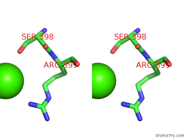
Calcium binding site 2 out of 2 in 1xhb
Go back to
Calcium binding site 2 out
of 2 in the The Crystal Structure of Udp-Galnac: Polypeptide Alpha-N- Acetylgalactosaminyltransferase-T1
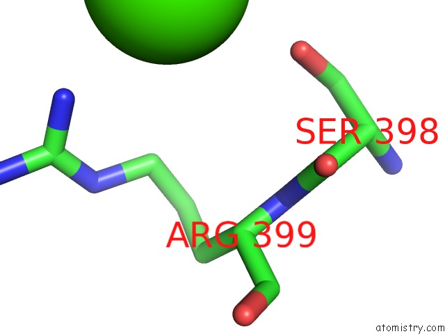
Mono view
Stereo pair view
Mono view
Stereo pair view
A full contact list of Calcium with other atoms in the Ca binding
site number 2 of The Crystal Structure of Udp-Galnac: Polypeptide Alpha-N- Acetylgalactosaminyltransferase-T1 within 5.0Å range:
|
Reference:
T.A.Fritz,
J.H.Hurley,
L.B.Trinh,
J.Shiloach,
L.A.Tabak.
The Beginnings of Mucin Biosynthesis: the Crystal Structure of Udp-Galnac:Polypeptide {Alpha}-N-Acetylgalactosaminyltransferase-T1 Proc.Natl.Acad.Sci.Usa V. 101 15307 2004.
ISSN: ISSN 0027-8424
PubMed: 15486088
DOI: 10.1073/PNAS.0405657101
Page generated: Fri Jul 12 07:32:25 2024
ISSN: ISSN 0027-8424
PubMed: 15486088
DOI: 10.1073/PNAS.0405657101
Last articles
Zn in 9J0NZn in 9J0O
Zn in 9J0P
Zn in 9FJX
Zn in 9EKB
Zn in 9C0F
Zn in 9CAH
Zn in 9CH0
Zn in 9CH3
Zn in 9CH1