Calcium »
PDB 1y1x-1ydy »
1y3g »
Calcium in PDB 1y3g: Crystal Structure of A Silanediol Protease Inhibitor Bound to Thermolysin
Enzymatic activity of Crystal Structure of A Silanediol Protease Inhibitor Bound to Thermolysin
All present enzymatic activity of Crystal Structure of A Silanediol Protease Inhibitor Bound to Thermolysin:
3.4.24.27;
3.4.24.27;
Protein crystallography data
The structure of Crystal Structure of A Silanediol Protease Inhibitor Bound to Thermolysin, PDB code: 1y3g
was solved by
D.H.Juers,
J.Kim,
B.W.Matthews,
S.M.Sieburth,
with X-Ray Crystallography technique. A brief refinement statistics is given in the table below:
| Resolution Low / High (Å) | 31.00 / 2.10 |
| Space group | P 61 2 2 |
| Cell size a, b, c (Å), α, β, γ (°) | 93.520, 93.520, 131.770, 90.00, 90.00, 120.00 |
| R / Rfree (%) | 15.5 / 22.5 |
Other elements in 1y3g:
The structure of Crystal Structure of A Silanediol Protease Inhibitor Bound to Thermolysin also contains other interesting chemical elements:
| Silicon | (Si) | 1 atom |
| Zinc | (Zn) | 1 atom |
Calcium Binding Sites:
The binding sites of Calcium atom in the Crystal Structure of A Silanediol Protease Inhibitor Bound to Thermolysin
(pdb code 1y3g). This binding sites where shown within
5.0 Angstroms radius around Calcium atom.
In total 4 binding sites of Calcium where determined in the Crystal Structure of A Silanediol Protease Inhibitor Bound to Thermolysin, PDB code: 1y3g:
Jump to Calcium binding site number: 1; 2; 3; 4;
In total 4 binding sites of Calcium where determined in the Crystal Structure of A Silanediol Protease Inhibitor Bound to Thermolysin, PDB code: 1y3g:
Jump to Calcium binding site number: 1; 2; 3; 4;
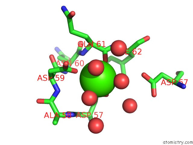
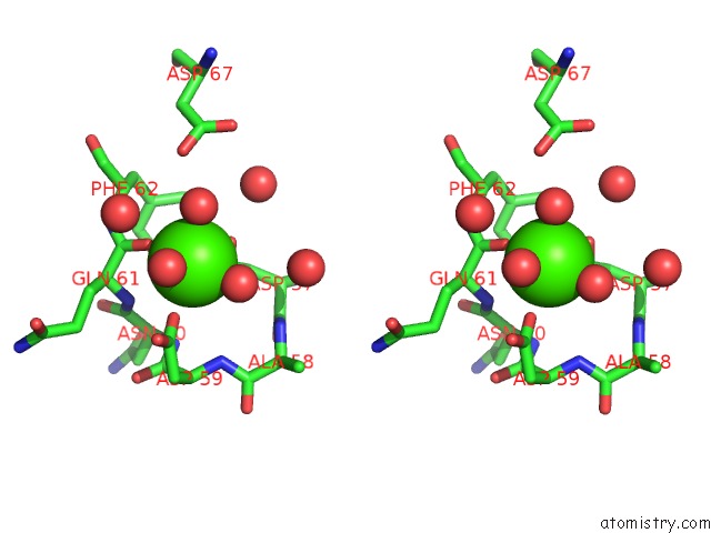
Calcium binding site 1 out of 4 in 1y3g
Go back to
Calcium binding site 1 out
of 4 in the Crystal Structure of A Silanediol Protease Inhibitor Bound to Thermolysin
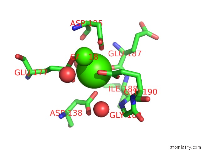
Mono view
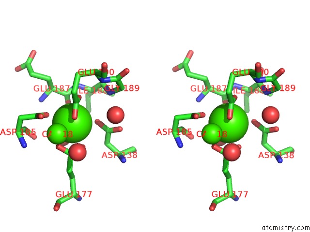
Stereo pair view
Mono view
Stereo pair view
A full contact list of Calcium with other atoms in the Ca binding
site number 1 of Crystal Structure of A Silanediol Protease Inhibitor Bound to Thermolysin within 5.0Å range:
|
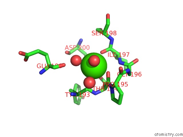
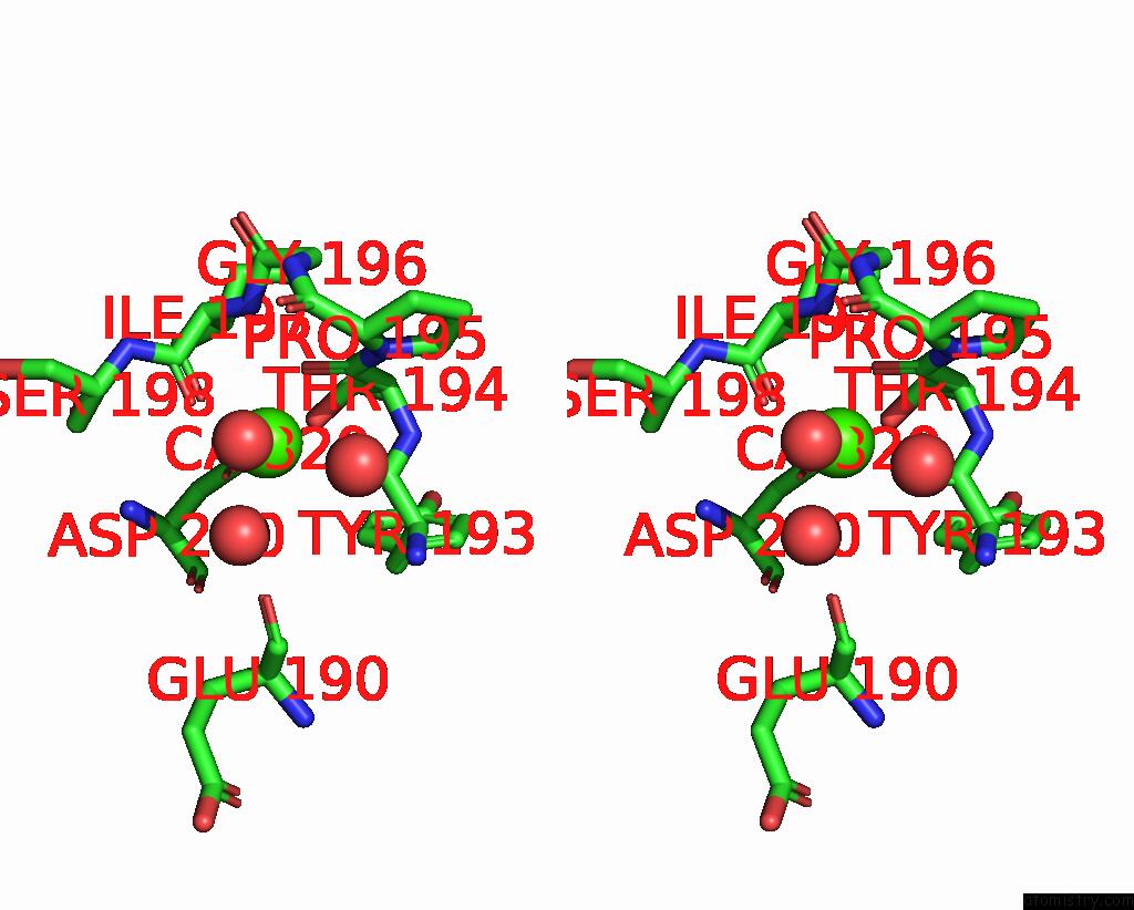
Calcium binding site 2 out of 4 in 1y3g
Go back to
Calcium binding site 2 out
of 4 in the Crystal Structure of A Silanediol Protease Inhibitor Bound to Thermolysin
Mono view
Stereo pair view
Mono view
Stereo pair view
A full contact list of Calcium with other atoms in the Ca binding
site number 2 of Crystal Structure of A Silanediol Protease Inhibitor Bound to Thermolysin within 5.0Å range:
|
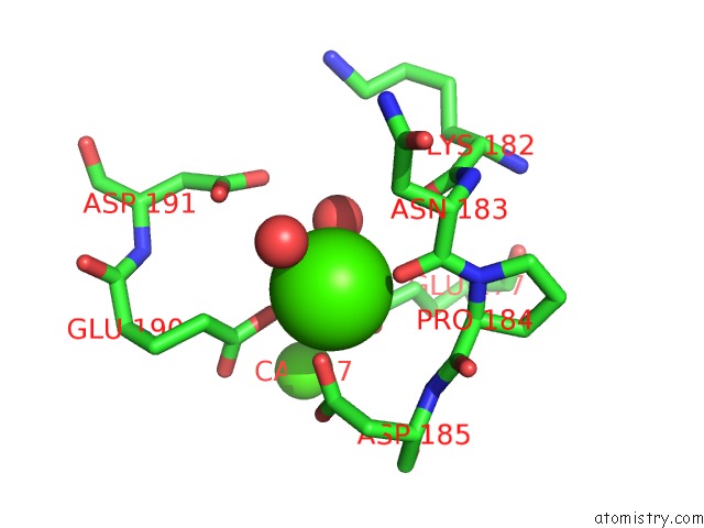
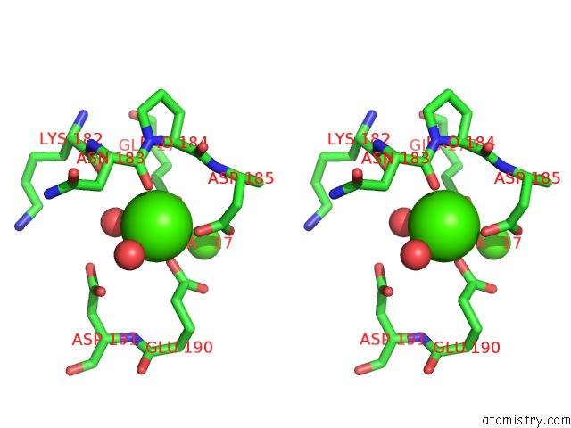
Calcium binding site 3 out of 4 in 1y3g
Go back to
Calcium binding site 3 out
of 4 in the Crystal Structure of A Silanediol Protease Inhibitor Bound to Thermolysin
Mono view
Stereo pair view
Mono view
Stereo pair view
A full contact list of Calcium with other atoms in the Ca binding
site number 3 of Crystal Structure of A Silanediol Protease Inhibitor Bound to Thermolysin within 5.0Å range:
|
Calcium binding site 4 out of 4 in 1y3g
Go back to
Calcium binding site 4 out
of 4 in the Crystal Structure of A Silanediol Protease Inhibitor Bound to Thermolysin
Mono view
Stereo pair view
Mono view
Stereo pair view
A full contact list of Calcium with other atoms in the Ca binding
site number 4 of Crystal Structure of A Silanediol Protease Inhibitor Bound to Thermolysin within 5.0Å range:
|
Reference:
D.H.Juers,
J.Kim,
B.W.Matthews,
S.M.Sieburth.
Structural Analysis of Silanediols As Transition-State-Analogue Inhibitors of the Benchmark Metalloprotease Thermolysin(,). Biochemistry V. 44 16524 2005.
ISSN: ISSN 0006-2960
PubMed: 16342943
DOI: 10.1021/BI051346V
Page generated: Fri Jul 12 07:57:35 2024
ISSN: ISSN 0006-2960
PubMed: 16342943
DOI: 10.1021/BI051346V
Last articles
Zn in 9JYWZn in 9IR4
Zn in 9IR3
Zn in 9GMX
Zn in 9GMW
Zn in 9JEJ
Zn in 9ERF
Zn in 9ERE
Zn in 9EGV
Zn in 9EGW