Calcium »
PDB 1y1x-1ydy »
1y64 »
Calcium in PDB 1y64: BNI1P Formin Homology 2 Domain Complexed with Atp-Actin
Protein crystallography data
The structure of BNI1P Formin Homology 2 Domain Complexed with Atp-Actin, PDB code: 1y64
was solved by
T.Otomo,
D.R.Tomchick,
C.Otomo,
S.C.Panchal,
M.Machius,
M.K.Rosen,
with X-Ray Crystallography technique. A brief refinement statistics is given in the table below:
| Resolution Low / High (Å) | 30.00 / 3.05 |
| Space group | C 1 2 1 |
| Cell size a, b, c (Å), α, β, γ (°) | 231.997, 56.229, 100.945, 90.00, 107.70, 90.00 |
| R / Rfree (%) | 28.9 / 31.3 |
Calcium Binding Sites:
The binding sites of Calcium atom in the BNI1P Formin Homology 2 Domain Complexed with Atp-Actin
(pdb code 1y64). This binding sites where shown within
5.0 Angstroms radius around Calcium atom.
In total only one binding site of Calcium was determined in the BNI1P Formin Homology 2 Domain Complexed with Atp-Actin, PDB code: 1y64:
In total only one binding site of Calcium was determined in the BNI1P Formin Homology 2 Domain Complexed with Atp-Actin, PDB code: 1y64:
Calcium binding site 1 out of 1 in 1y64
Go back to
Calcium binding site 1 out
of 1 in the BNI1P Formin Homology 2 Domain Complexed with Atp-Actin

Mono view
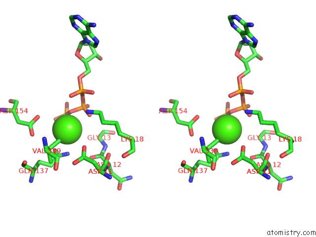
Stereo pair view
Mono view
Stereo pair view
A full contact list of Calcium with other atoms in the Ca binding
site number 1 of BNI1P Formin Homology 2 Domain Complexed with Atp-Actin within 5.0Å range:
|
Reference:
T.Otomo,
D.R.Tomchick,
C.Otomo,
S.C.Panchal,
M.Machius,
M.K.Rosen.
Structural Basis of Actin Filament Nucleation and Processive Capping By A Formin Homology 2 Domain Nature V. 433 488 2005.
ISSN: ISSN 0028-0836
PubMed: 15635372
DOI: 10.1038/NATURE03251
Page generated: Fri Jul 12 08:00:37 2024
ISSN: ISSN 0028-0836
PubMed: 15635372
DOI: 10.1038/NATURE03251
Last articles
Zn in 9J0NZn in 9J0O
Zn in 9J0P
Zn in 9FJX
Zn in 9EKB
Zn in 9C0F
Zn in 9CAH
Zn in 9CH0
Zn in 9CH3
Zn in 9CH1