Calcium »
PDB 1y1x-1ydy »
1y6o »
Calcium in PDB 1y6o: Crystal Structure of Disulfide Engineered Porcine Pancreatic Phospholipase A2 to Group-X Isozyme in Complex with Inhibitor MJ33 and Phosphate Ions
Enzymatic activity of Crystal Structure of Disulfide Engineered Porcine Pancreatic Phospholipase A2 to Group-X Isozyme in Complex with Inhibitor MJ33 and Phosphate Ions
All present enzymatic activity of Crystal Structure of Disulfide Engineered Porcine Pancreatic Phospholipase A2 to Group-X Isozyme in Complex with Inhibitor MJ33 and Phosphate Ions:
3.1.1.4;
3.1.1.4;
Protein crystallography data
The structure of Crystal Structure of Disulfide Engineered Porcine Pancreatic Phospholipase A2 to Group-X Isozyme in Complex with Inhibitor MJ33 and Phosphate Ions, PDB code: 1y6o
was solved by
B.Z.Yu,
Y.H.Pan,
M.J.W.Jassen,
B.J.Bahnson,
M.K.Jain,
with X-Ray Crystallography technique. A brief refinement statistics is given in the table below:
| Resolution Low / High (Å) | 28.64 / 2.00 |
| Space group | P 1 21 1 |
| Cell size a, b, c (Å), α, β, γ (°) | 38.534, 85.921, 38.554, 90.00, 94.76, 90.00 |
| R / Rfree (%) | 19.6 / 24.5 |
Other elements in 1y6o:
The structure of Crystal Structure of Disulfide Engineered Porcine Pancreatic Phospholipase A2 to Group-X Isozyme in Complex with Inhibitor MJ33 and Phosphate Ions also contains other interesting chemical elements:
| Fluorine | (F) | 6 atoms |
Calcium Binding Sites:
The binding sites of Calcium atom in the Crystal Structure of Disulfide Engineered Porcine Pancreatic Phospholipase A2 to Group-X Isozyme in Complex with Inhibitor MJ33 and Phosphate Ions
(pdb code 1y6o). This binding sites where shown within
5.0 Angstroms radius around Calcium atom.
In total 2 binding sites of Calcium where determined in the Crystal Structure of Disulfide Engineered Porcine Pancreatic Phospholipase A2 to Group-X Isozyme in Complex with Inhibitor MJ33 and Phosphate Ions, PDB code: 1y6o:
Jump to Calcium binding site number: 1; 2;
In total 2 binding sites of Calcium where determined in the Crystal Structure of Disulfide Engineered Porcine Pancreatic Phospholipase A2 to Group-X Isozyme in Complex with Inhibitor MJ33 and Phosphate Ions, PDB code: 1y6o:
Jump to Calcium binding site number: 1; 2;
Calcium binding site 1 out of 2 in 1y6o
Go back to
Calcium binding site 1 out
of 2 in the Crystal Structure of Disulfide Engineered Porcine Pancreatic Phospholipase A2 to Group-X Isozyme in Complex with Inhibitor MJ33 and Phosphate Ions
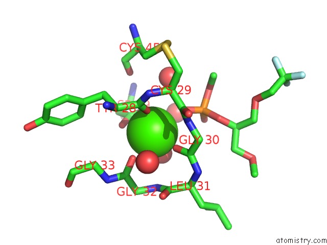
Mono view
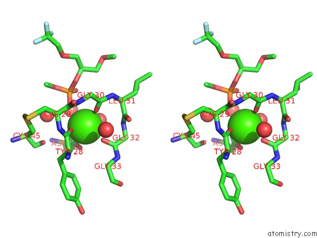
Stereo pair view
Mono view
Stereo pair view
A full contact list of Calcium with other atoms in the Ca binding
site number 1 of Crystal Structure of Disulfide Engineered Porcine Pancreatic Phospholipase A2 to Group-X Isozyme in Complex with Inhibitor MJ33 and Phosphate Ions within 5.0Å range:
|
Calcium binding site 2 out of 2 in 1y6o
Go back to
Calcium binding site 2 out
of 2 in the Crystal Structure of Disulfide Engineered Porcine Pancreatic Phospholipase A2 to Group-X Isozyme in Complex with Inhibitor MJ33 and Phosphate Ions
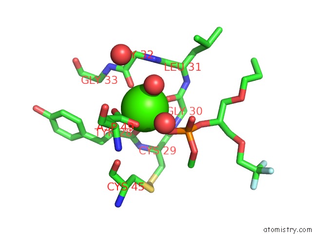
Mono view
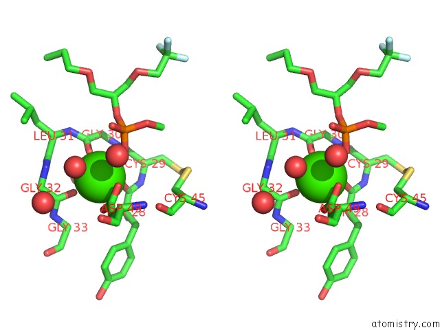
Stereo pair view
Mono view
Stereo pair view
A full contact list of Calcium with other atoms in the Ca binding
site number 2 of Crystal Structure of Disulfide Engineered Porcine Pancreatic Phospholipase A2 to Group-X Isozyme in Complex with Inhibitor MJ33 and Phosphate Ions within 5.0Å range:
|
Reference:
B.Z.Yu,
Y.H.Pan,
M.J.W.Janssen,
B.J.Bahnson,
M.K.Jain.
Kinetic and Structural Properties of Disulfide Engineered Phospholipase A(2): Insight Into the Role of Disulfide Bonding Patterns. Biochemistry V. 44 3369 2005.
ISSN: ISSN 0006-2960
PubMed: 15736947
DOI: 10.1021/BI0482147
Page generated: Fri Jul 12 08:00:47 2024
ISSN: ISSN 0006-2960
PubMed: 15736947
DOI: 10.1021/BI0482147
Last articles
Zn in 9MJ5Zn in 9HNW
Zn in 9G0L
Zn in 9FNE
Zn in 9DZN
Zn in 9E0I
Zn in 9D32
Zn in 9DAK
Zn in 8ZXC
Zn in 8ZUF