Calcium »
PDB 1yvu-1zez »
1z0b »
Calcium in PDB 1z0b: Crystal Structure of A. Fulgidus Lon Proteolytic Domain E506A Mutant
Enzymatic activity of Crystal Structure of A. Fulgidus Lon Proteolytic Domain E506A Mutant
All present enzymatic activity of Crystal Structure of A. Fulgidus Lon Proteolytic Domain E506A Mutant:
3.4.21.53;
3.4.21.53;
Protein crystallography data
The structure of Crystal Structure of A. Fulgidus Lon Proteolytic Domain E506A Mutant, PDB code: 1z0b
was solved by
I.Botos,
E.E.Melnikov,
S.Cherry,
S.Kozlov,
O.V.Makhovskaya,
J.E.Tropea,
A.Gustchina,
T.V.Rotanova,
A.Wlodawer,
with X-Ray Crystallography technique. A brief refinement statistics is given in the table below:
| Resolution Low / High (Å) | 20.00 / 1.55 |
| Space group | P 65 |
| Cell size a, b, c (Å), α, β, γ (°) | 83.260, 83.260, 41.170, 90.00, 90.00, 120.00 |
| R / Rfree (%) | 18 / 20.7 |
Calcium Binding Sites:
The binding sites of Calcium atom in the Crystal Structure of A. Fulgidus Lon Proteolytic Domain E506A Mutant
(pdb code 1z0b). This binding sites where shown within
5.0 Angstroms radius around Calcium atom.
In total 2 binding sites of Calcium where determined in the Crystal Structure of A. Fulgidus Lon Proteolytic Domain E506A Mutant, PDB code: 1z0b:
Jump to Calcium binding site number: 1; 2;
In total 2 binding sites of Calcium where determined in the Crystal Structure of A. Fulgidus Lon Proteolytic Domain E506A Mutant, PDB code: 1z0b:
Jump to Calcium binding site number: 1; 2;
Calcium binding site 1 out of 2 in 1z0b
Go back to
Calcium binding site 1 out
of 2 in the Crystal Structure of A. Fulgidus Lon Proteolytic Domain E506A Mutant
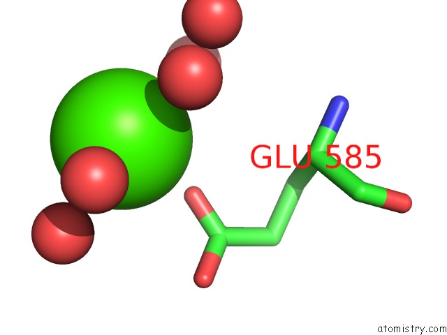
Mono view
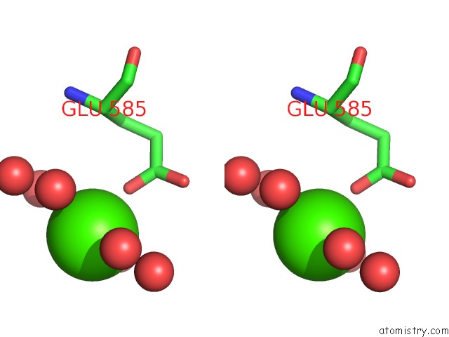
Stereo pair view
Mono view
Stereo pair view
A full contact list of Calcium with other atoms in the Ca binding
site number 1 of Crystal Structure of A. Fulgidus Lon Proteolytic Domain E506A Mutant within 5.0Å range:
|
Calcium binding site 2 out of 2 in 1z0b
Go back to
Calcium binding site 2 out
of 2 in the Crystal Structure of A. Fulgidus Lon Proteolytic Domain E506A Mutant
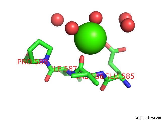
Mono view
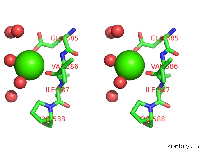
Stereo pair view
Mono view
Stereo pair view
A full contact list of Calcium with other atoms in the Ca binding
site number 2 of Crystal Structure of A. Fulgidus Lon Proteolytic Domain E506A Mutant within 5.0Å range:
|
Reference:
I.Botos,
E.E.Melnikov,
S.Cherry,
S.Kozlov,
O.V.Makhovskaya,
J.E.Tropea,
A.Gustchina,
T.V.Rotanova,
A.Wlodawer.
Atomic-Resolution Crystal Structure of the Proteolytic Domain of Archaeoglobus Fulgidus Lon Reveals the Conformational Variability in the Active Sites of Lon Proteases J.Mol.Biol. V. 351 144 2005.
ISSN: ISSN 0022-2836
PubMed: 16002085
DOI: 10.1016/J.JMB.2005.06.008
Page generated: Fri Jul 12 08:18:34 2024
ISSN: ISSN 0022-2836
PubMed: 16002085
DOI: 10.1016/J.JMB.2005.06.008
Last articles
Zn in 9J0NZn in 9J0O
Zn in 9J0P
Zn in 9FJX
Zn in 9EKB
Zn in 9C0F
Zn in 9CAH
Zn in 9CH0
Zn in 9CH3
Zn in 9CH1