Calcium »
PDB 1yvu-1zez »
1z29 »
Calcium in PDB 1z29: Crystal Structures of SULT1A2 and SULT1A1*3: Implications in the Bioactivation of N-Hydroxy-2-Acetylamino Fluorine (Oh-Aaf)
Enzymatic activity of Crystal Structures of SULT1A2 and SULT1A1*3: Implications in the Bioactivation of N-Hydroxy-2-Acetylamino Fluorine (Oh-Aaf)
All present enzymatic activity of Crystal Structures of SULT1A2 and SULT1A1*3: Implications in the Bioactivation of N-Hydroxy-2-Acetylamino Fluorine (Oh-Aaf):
2.8.2.1;
2.8.2.1;
Protein crystallography data
The structure of Crystal Structures of SULT1A2 and SULT1A1*3: Implications in the Bioactivation of N-Hydroxy-2-Acetylamino Fluorine (Oh-Aaf), PDB code: 1z29
was solved by
J.Lu,
H.Li,
M.C.Liu,
J.Zhang,
M.Li,
X.An,
W.Chang,
with X-Ray Crystallography technique. A brief refinement statistics is given in the table below:
| Resolution Low / High (Å) | 50.00 / 2.40 |
| Space group | C 1 2 1 |
| Cell size a, b, c (Å), α, β, γ (°) | 58.398, 65.399, 86.517, 90.00, 103.69, 90.00 |
| R / Rfree (%) | 21.6 / 25.3 |
Calcium Binding Sites:
The binding sites of Calcium atom in the Crystal Structures of SULT1A2 and SULT1A1*3: Implications in the Bioactivation of N-Hydroxy-2-Acetylamino Fluorine (Oh-Aaf)
(pdb code 1z29). This binding sites where shown within
5.0 Angstroms radius around Calcium atom.
In total only one binding site of Calcium was determined in the Crystal Structures of SULT1A2 and SULT1A1*3: Implications in the Bioactivation of N-Hydroxy-2-Acetylamino Fluorine (Oh-Aaf), PDB code: 1z29:
In total only one binding site of Calcium was determined in the Crystal Structures of SULT1A2 and SULT1A1*3: Implications in the Bioactivation of N-Hydroxy-2-Acetylamino Fluorine (Oh-Aaf), PDB code: 1z29:
Calcium binding site 1 out of 1 in 1z29
Go back to
Calcium binding site 1 out
of 1 in the Crystal Structures of SULT1A2 and SULT1A1*3: Implications in the Bioactivation of N-Hydroxy-2-Acetylamino Fluorine (Oh-Aaf)
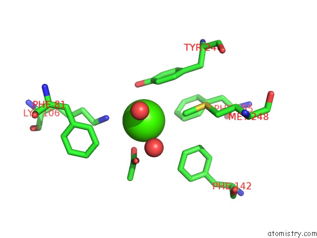
Mono view
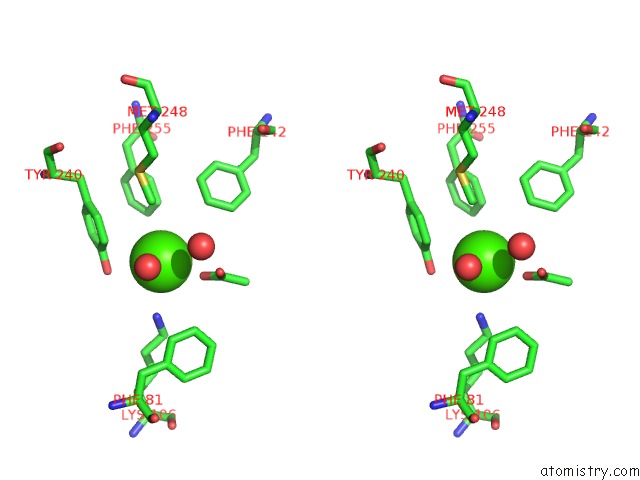
Stereo pair view
Mono view
Stereo pair view
A full contact list of Calcium with other atoms in the Ca binding
site number 1 of Crystal Structures of SULT1A2 and SULT1A1*3: Implications in the Bioactivation of N-Hydroxy-2-Acetylamino Fluorine (Oh-Aaf) within 5.0Å range:
|
Reference:
J.Lu,
H.Li,
J.Zhang,
M.Li,
M.Y.Liu,
X.An,
M.C.Liu,
W.Chang.
Crystal Structures of SULT1A2 and SULT1A1 *3: Insights Into the Substrate Inhibition and the Role of TYR149 in SULT1A2. Biochem.Biophys.Res.Commun. V. 396 429 2010.
ISSN: ISSN 0006-291X
PubMed: 20417180
DOI: 10.1016/J.BBRC.2010.04.109
Page generated: Tue Jul 8 04:00:24 2025
ISSN: ISSN 0006-291X
PubMed: 20417180
DOI: 10.1016/J.BBRC.2010.04.109
Last articles
F in 4J0PF in 4IZW
F in 4IW8
F in 4IZT
F in 4IWF
F in 4IXE
F in 4IW6
F in 4IVW
F in 4IVY
F in 4IVO