Calcium »
PDB 1yvu-1zez »
1z3s »
Calcium in PDB 1z3s: Angiopoietin-2 Receptor Binding Domain
Protein crystallography data
The structure of Angiopoietin-2 Receptor Binding Domain, PDB code: 1z3s
was solved by
W.A.Barton,
D.Tzvetkova,
D.B.Nikolov,
with X-Ray Crystallography technique. A brief refinement statistics is given in the table below:
| Resolution Low / High (Å) | 8.00 / 2.35 |
| Space group | C 1 2 1 |
| Cell size a, b, c (Å), α, β, γ (°) | 74.190, 136.448, 47.732, 90.00, 93.43, 90.00 |
| R / Rfree (%) | 22.9 / 28.7 |
Calcium Binding Sites:
The binding sites of Calcium atom in the Angiopoietin-2 Receptor Binding Domain
(pdb code 1z3s). This binding sites where shown within
5.0 Angstroms radius around Calcium atom.
In total 2 binding sites of Calcium where determined in the Angiopoietin-2 Receptor Binding Domain, PDB code: 1z3s:
Jump to Calcium binding site number: 1; 2;
In total 2 binding sites of Calcium where determined in the Angiopoietin-2 Receptor Binding Domain, PDB code: 1z3s:
Jump to Calcium binding site number: 1; 2;
Calcium binding site 1 out of 2 in 1z3s
Go back to
Calcium binding site 1 out
of 2 in the Angiopoietin-2 Receptor Binding Domain
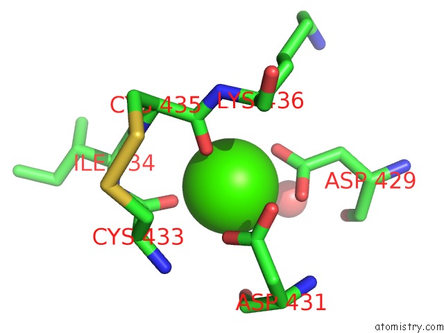
Mono view
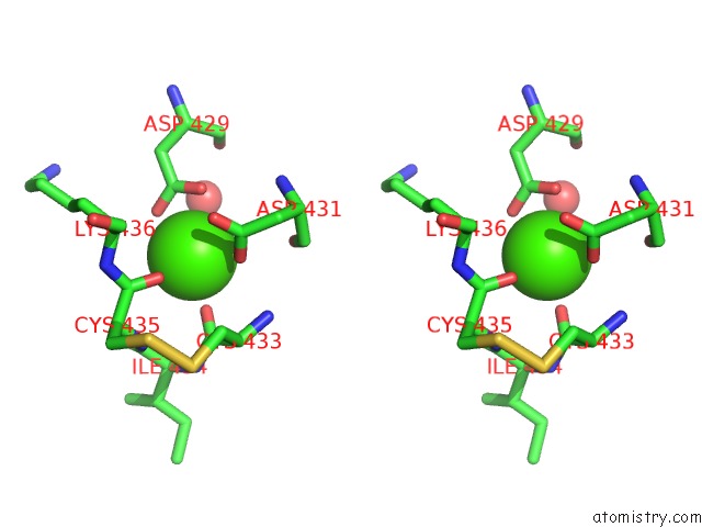
Stereo pair view
Mono view
Stereo pair view
A full contact list of Calcium with other atoms in the Ca binding
site number 1 of Angiopoietin-2 Receptor Binding Domain within 5.0Å range:
|
Calcium binding site 2 out of 2 in 1z3s
Go back to
Calcium binding site 2 out
of 2 in the Angiopoietin-2 Receptor Binding Domain
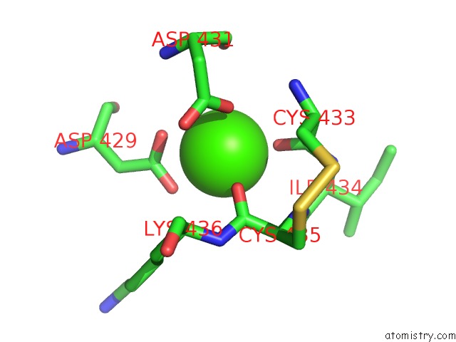
Mono view
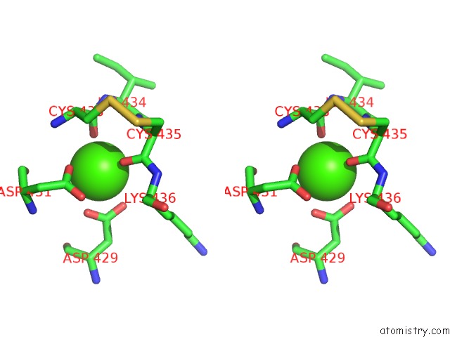
Stereo pair view
Mono view
Stereo pair view
A full contact list of Calcium with other atoms in the Ca binding
site number 2 of Angiopoietin-2 Receptor Binding Domain within 5.0Å range:
|
Reference:
W.A.Barton,
D.Tzvetkova,
D.B.Nikolov.
Structure of the Angiopoietin-2 Receptor Binding Domain and Identification of Surfaces Involved in TIE2 Recognition. Structure V. 13 825 2005.
ISSN: ISSN 0969-2126
PubMed: 15893672
DOI: 10.1016/J.STR.2005.03.009
Page generated: Fri Jul 12 08:19:38 2024
ISSN: ISSN 0969-2126
PubMed: 15893672
DOI: 10.1016/J.STR.2005.03.009
Last articles
Zn in 9J0NZn in 9J0O
Zn in 9J0P
Zn in 9FJX
Zn in 9EKB
Zn in 9C0F
Zn in 9CAH
Zn in 9CH0
Zn in 9CH3
Zn in 9CH1