Calcium »
PDB 1yvu-1zez »
1zdp »
Calcium in PDB 1zdp: Crystal Structure Analysis of Thermolysin Complexed with the Inhibitor (S)-Thiorphan
Enzymatic activity of Crystal Structure Analysis of Thermolysin Complexed with the Inhibitor (S)-Thiorphan
All present enzymatic activity of Crystal Structure Analysis of Thermolysin Complexed with the Inhibitor (S)-Thiorphan:
3.4.24.27;
3.4.24.27;
Protein crystallography data
The structure of Crystal Structure Analysis of Thermolysin Complexed with the Inhibitor (S)-Thiorphan, PDB code: 1zdp
was solved by
S.L.Roderick,
M.C.Fournie-Zaluski,
B.P.Roques,
B.W.Matthews,
with X-Ray Crystallography technique. A brief refinement statistics is given in the table below:
| Resolution Low / High (Å) | 10.00 / 1.70 |
| Space group | P 61 2 2 |
| Cell size a, b, c (Å), α, β, γ (°) | 94.000, 94.000, 132.100, 90.00, 90.00, 120.00 |
| R / Rfree (%) | 18.3 / n/a |
Other elements in 1zdp:
The structure of Crystal Structure Analysis of Thermolysin Complexed with the Inhibitor (S)-Thiorphan also contains other interesting chemical elements:
| Zinc | (Zn) | 1 atom |
Calcium Binding Sites:
The binding sites of Calcium atom in the Crystal Structure Analysis of Thermolysin Complexed with the Inhibitor (S)-Thiorphan
(pdb code 1zdp). This binding sites where shown within
5.0 Angstroms radius around Calcium atom.
In total 4 binding sites of Calcium where determined in the Crystal Structure Analysis of Thermolysin Complexed with the Inhibitor (S)-Thiorphan, PDB code: 1zdp:
Jump to Calcium binding site number: 1; 2; 3; 4;
In total 4 binding sites of Calcium where determined in the Crystal Structure Analysis of Thermolysin Complexed with the Inhibitor (S)-Thiorphan, PDB code: 1zdp:
Jump to Calcium binding site number: 1; 2; 3; 4;
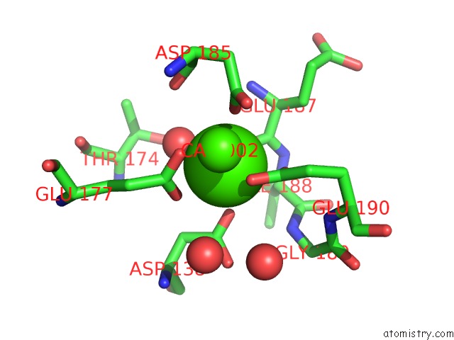
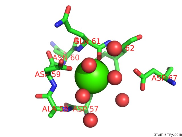
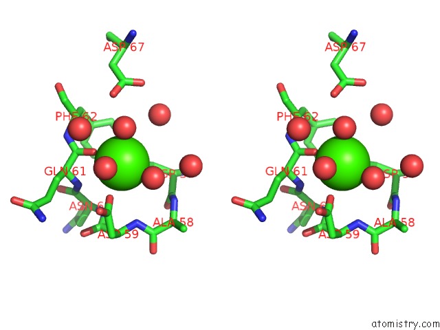
Calcium binding site 1 out of 4 in 1zdp
Go back to
Calcium binding site 1 out
of 4 in the Crystal Structure Analysis of Thermolysin Complexed with the Inhibitor (S)-Thiorphan
Mono view
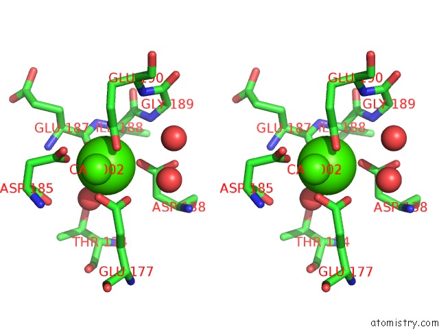
Stereo pair view
Mono view
Stereo pair view
A full contact list of Calcium with other atoms in the Ca binding
site number 1 of Crystal Structure Analysis of Thermolysin Complexed with the Inhibitor (S)-Thiorphan within 5.0Å range:
|
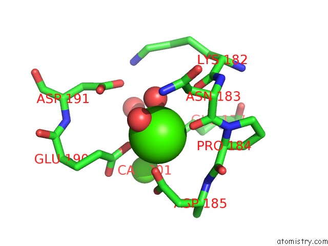
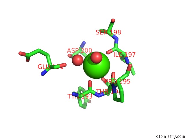
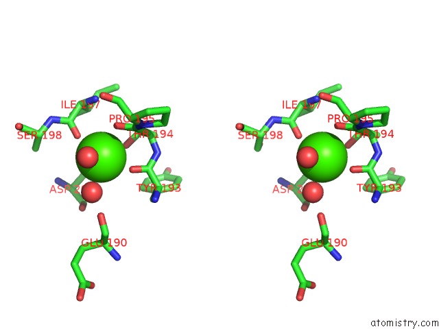
Calcium binding site 2 out of 4 in 1zdp
Go back to
Calcium binding site 2 out
of 4 in the Crystal Structure Analysis of Thermolysin Complexed with the Inhibitor (S)-Thiorphan
Mono view
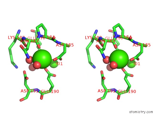
Stereo pair view
Mono view
Stereo pair view
A full contact list of Calcium with other atoms in the Ca binding
site number 2 of Crystal Structure Analysis of Thermolysin Complexed with the Inhibitor (S)-Thiorphan within 5.0Å range:
|
Calcium binding site 3 out of 4 in 1zdp
Go back to
Calcium binding site 3 out
of 4 in the Crystal Structure Analysis of Thermolysin Complexed with the Inhibitor (S)-Thiorphan
Mono view
Stereo pair view
Mono view
Stereo pair view
A full contact list of Calcium with other atoms in the Ca binding
site number 3 of Crystal Structure Analysis of Thermolysin Complexed with the Inhibitor (S)-Thiorphan within 5.0Å range:
|
Calcium binding site 4 out of 4 in 1zdp
Go back to
Calcium binding site 4 out
of 4 in the Crystal Structure Analysis of Thermolysin Complexed with the Inhibitor (S)-Thiorphan
Mono view
Stereo pair view
Mono view
Stereo pair view
A full contact list of Calcium with other atoms in the Ca binding
site number 4 of Crystal Structure Analysis of Thermolysin Complexed with the Inhibitor (S)-Thiorphan within 5.0Å range:
|
Reference:
S.L.Roderick,
M.C.Fournie-Zaluski,
B.P.Roques,
B.W.Matthews.
Thiorphan and Retro-Thiorphan Display Equivalent Interactions When Bound to Crystalline Thermolysin Biochemistry V. 28 1493 1989.
ISSN: ISSN 0006-2960
PubMed: 2719912
DOI: 10.1021/BI00430A011
Page generated: Tue Jul 8 04:03:15 2025
ISSN: ISSN 0006-2960
PubMed: 2719912
DOI: 10.1021/BI00430A011
Last articles
Cl in 5KMDCl in 5KMB
Cl in 5KMA
Cl in 5KM9
Cl in 5KM8
Cl in 5KM5
Cl in 5KKI
Cl in 5KM2
Cl in 5KLV
Cl in 5KKY