Calcium »
PDB 1zf0-2a2z »
1zfb »
Calcium in PDB 1zfb: Ggc Duplex B-Dna
Protein crystallography data
The structure of Ggc Duplex B-Dna, PDB code: 1zfb
was solved by
F.A.Hays,
A.T.Teegarden,
Z.J.R.Jones,
M.Harms,
D.Raup,
J.Watson,
E.Cavaliere,
P.S.Ho,
with X-Ray Crystallography technique. A brief refinement statistics is given in the table below:
| Resolution Low / High (Å) | 16.35 / 1.65 |
| Space group | H 3 |
| Cell size a, b, c (Å), α, β, γ (°) | 53.640, 53.640, 44.820, 90.00, 90.00, 120.00 |
| R / Rfree (%) | 23.3 / 28.3 |
Calcium Binding Sites:
The binding sites of Calcium atom in the Ggc Duplex B-Dna
(pdb code 1zfb). This binding sites where shown within
5.0 Angstroms radius around Calcium atom.
In total 2 binding sites of Calcium where determined in the Ggc Duplex B-Dna, PDB code: 1zfb:
Jump to Calcium binding site number: 1; 2;
In total 2 binding sites of Calcium where determined in the Ggc Duplex B-Dna, PDB code: 1zfb:
Jump to Calcium binding site number: 1; 2;
Calcium binding site 1 out of 2 in 1zfb
Go back to
Calcium binding site 1 out
of 2 in the Ggc Duplex B-Dna
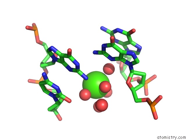
Mono view
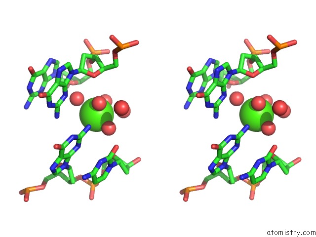
Stereo pair view
Mono view
Stereo pair view
A full contact list of Calcium with other atoms in the Ca binding
site number 1 of Ggc Duplex B-Dna within 5.0Å range:
|
Calcium binding site 2 out of 2 in 1zfb
Go back to
Calcium binding site 2 out
of 2 in the Ggc Duplex B-Dna
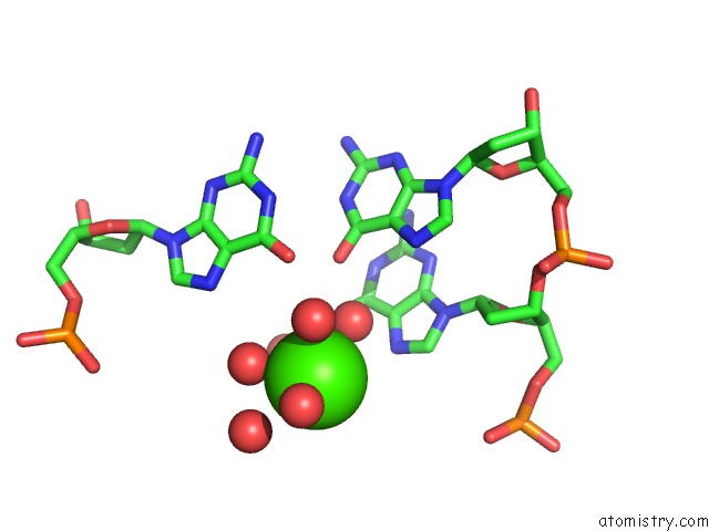
Mono view
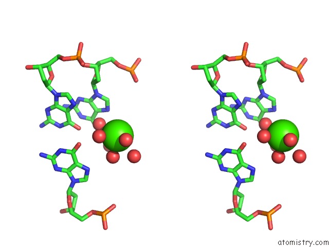
Stereo pair view
Mono view
Stereo pair view
A full contact list of Calcium with other atoms in the Ca binding
site number 2 of Ggc Duplex B-Dna within 5.0Å range:
|
Reference:
F.A.Hays,
A.Teegarden,
Z.J.Jones,
M.Harms,
D.Raup,
J.Watson,
E.Cavaliere,
P.S.Ho.
How Sequence Defines Structure: A Crystallographic Map of Dna Structure and Conformation. Proc.Natl.Acad.Sci.Usa V. 102 7157 2005.
ISSN: ISSN 0027-8424
PubMed: 15870206
DOI: 10.1073/PNAS.0409455102
Page generated: Fri Jul 12 08:24:37 2024
ISSN: ISSN 0027-8424
PubMed: 15870206
DOI: 10.1073/PNAS.0409455102
Last articles
Zn in 9MJ5Zn in 9HNW
Zn in 9G0L
Zn in 9FNE
Zn in 9DZN
Zn in 9E0I
Zn in 9D32
Zn in 9DAK
Zn in 8ZXC
Zn in 8ZUF