Calcium »
PDB 1zf0-2a2z »
1zot »
Calcium in PDB 1zot: Crystal Structure Analysis of the Cyaa/C-Cam with Pmeapp
Enzymatic activity of Crystal Structure Analysis of the Cyaa/C-Cam with Pmeapp
All present enzymatic activity of Crystal Structure Analysis of the Cyaa/C-Cam with Pmeapp:
4.6.1.1;
4.6.1.1;
Protein crystallography data
The structure of Crystal Structure Analysis of the Cyaa/C-Cam with Pmeapp, PDB code: 1zot
was solved by
Q.Guo,
W.J.Tang,
with X-Ray Crystallography technique. A brief refinement statistics is given in the table below:
| Resolution Low / High (Å) | 34.52 / 2.20 |
| Space group | P 41 21 2 |
| Cell size a, b, c (Å), α, β, γ (°) | 79.674, 79.674, 139.339, 90.00, 90.00, 90.00 |
| R / Rfree (%) | 25.2 / 29.1 |
Other elements in 1zot:
The structure of Crystal Structure Analysis of the Cyaa/C-Cam with Pmeapp also contains other interesting chemical elements:
| Magnesium | (Mg) | 3 atoms |
Calcium Binding Sites:
The binding sites of Calcium atom in the Crystal Structure Analysis of the Cyaa/C-Cam with Pmeapp
(pdb code 1zot). This binding sites where shown within
5.0 Angstroms radius around Calcium atom.
In total 2 binding sites of Calcium where determined in the Crystal Structure Analysis of the Cyaa/C-Cam with Pmeapp, PDB code: 1zot:
Jump to Calcium binding site number: 1; 2;
In total 2 binding sites of Calcium where determined in the Crystal Structure Analysis of the Cyaa/C-Cam with Pmeapp, PDB code: 1zot:
Jump to Calcium binding site number: 1; 2;
Calcium binding site 1 out of 2 in 1zot
Go back to
Calcium binding site 1 out
of 2 in the Crystal Structure Analysis of the Cyaa/C-Cam with Pmeapp

Mono view
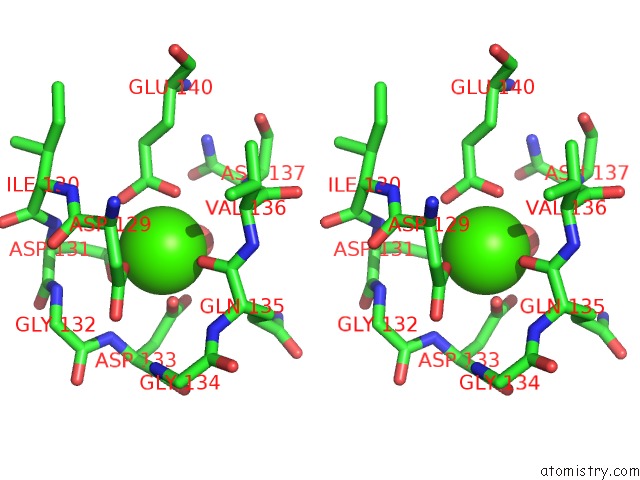
Stereo pair view
Mono view
Stereo pair view
A full contact list of Calcium with other atoms in the Ca binding
site number 1 of Crystal Structure Analysis of the Cyaa/C-Cam with Pmeapp within 5.0Å range:
|
Calcium binding site 2 out of 2 in 1zot
Go back to
Calcium binding site 2 out
of 2 in the Crystal Structure Analysis of the Cyaa/C-Cam with Pmeapp

Mono view
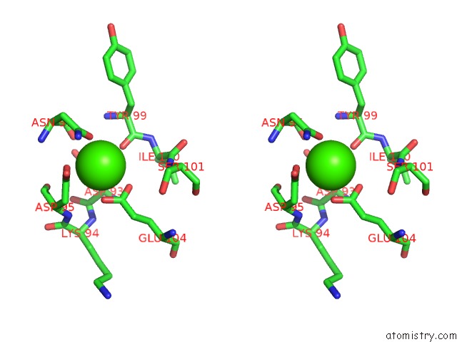
Stereo pair view
Mono view
Stereo pair view
A full contact list of Calcium with other atoms in the Ca binding
site number 2 of Crystal Structure Analysis of the Cyaa/C-Cam with Pmeapp within 5.0Å range:
|
Reference:
Q.Guo,
Y.Shen,
Y.S.Lee,
C.S.Gibbs,
M.Mrksich,
W.J.Tang.
Structural Basis For the Interaction of Bordetella Pertussis Adenylyl Cyclase Toxin with Calmodulin. Embo J. V. 24 3190 2005.
ISSN: ISSN 0261-4189
PubMed: 16138079
DOI: 10.1038/SJ.EMBOJ.7600800
Page generated: Fri Jul 12 08:26:49 2024
ISSN: ISSN 0261-4189
PubMed: 16138079
DOI: 10.1038/SJ.EMBOJ.7600800
Last articles
Zn in 9J0NZn in 9J0O
Zn in 9J0P
Zn in 9FJX
Zn in 9EKB
Zn in 9C0F
Zn in 9CAH
Zn in 9CH0
Zn in 9CH3
Zn in 9CH1