Calcium »
PDB 1zf0-2a2z »
1zud »
Calcium in PDB 1zud: Structure of This-Thif Protein Complex
Protein crystallography data
The structure of Structure of This-Thif Protein Complex, PDB code: 1zud
was solved by
S.E.Ealick,
C.Lehmann,
with X-Ray Crystallography technique. A brief refinement statistics is given in the table below:
| Resolution Low / High (Å) | 24.23 / 1.98 |
| Space group | P 21 21 21 |
| Cell size a, b, c (Å), α, β, γ (°) | 49.505, 111.171, 114.153, 90.00, 90.00, 90.00 |
| R / Rfree (%) | 17.4 / 22.9 |
Other elements in 1zud:
The structure of Structure of This-Thif Protein Complex also contains other interesting chemical elements:
| Zinc | (Zn) | 2 atoms |
| Sodium | (Na) | 2 atoms |
Calcium Binding Sites:
The binding sites of Calcium atom in the Structure of This-Thif Protein Complex
(pdb code 1zud). This binding sites where shown within
5.0 Angstroms radius around Calcium atom.
In total 2 binding sites of Calcium where determined in the Structure of This-Thif Protein Complex, PDB code: 1zud:
Jump to Calcium binding site number: 1; 2;
In total 2 binding sites of Calcium where determined in the Structure of This-Thif Protein Complex, PDB code: 1zud:
Jump to Calcium binding site number: 1; 2;
Calcium binding site 1 out of 2 in 1zud
Go back to
Calcium binding site 1 out
of 2 in the Structure of This-Thif Protein Complex
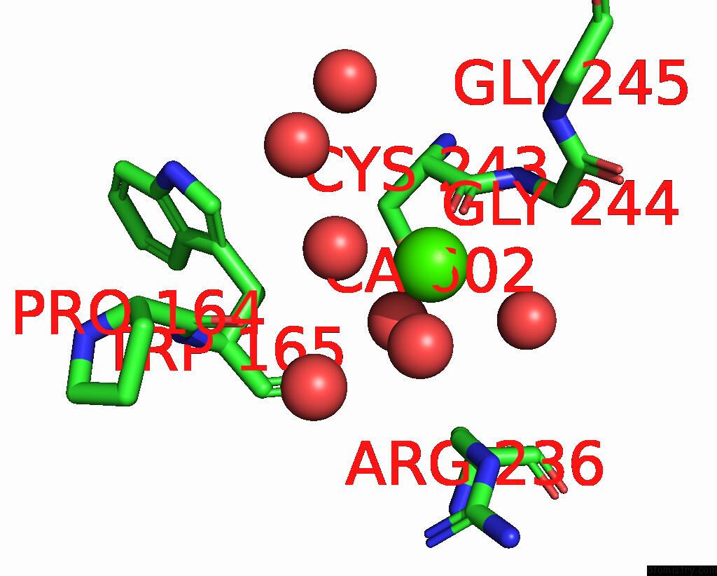
Mono view
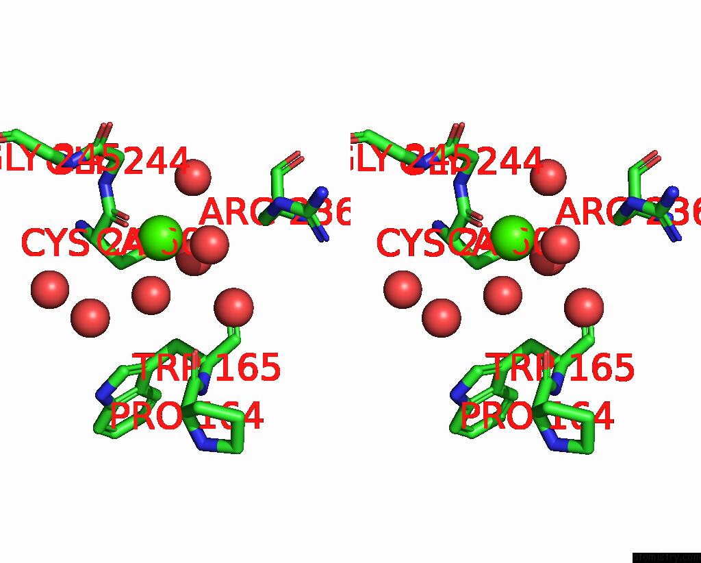
Stereo pair view
Mono view
Stereo pair view
A full contact list of Calcium with other atoms in the Ca binding
site number 1 of Structure of This-Thif Protein Complex within 5.0Å range:
|
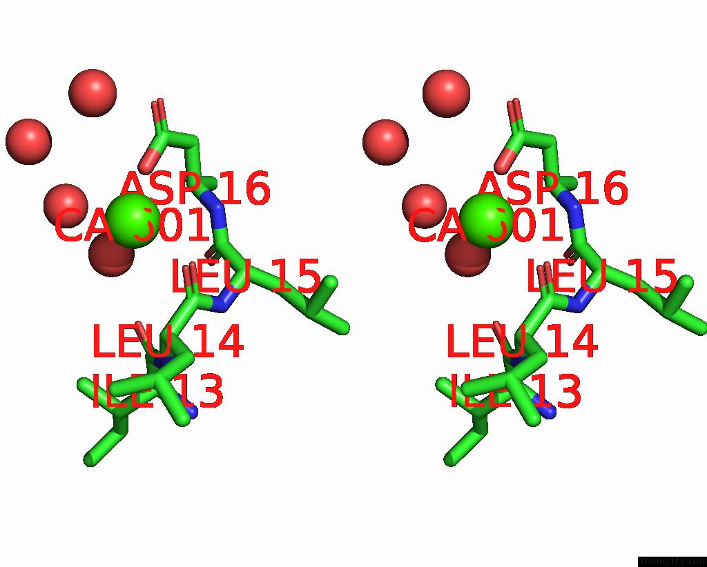
Calcium binding site 2 out of 2 in 1zud
Go back to
Calcium binding site 2 out
of 2 in the Structure of This-Thif Protein Complex

Mono view
Stereo pair view
Mono view
Stereo pair view
A full contact list of Calcium with other atoms in the Ca binding
site number 2 of Structure of This-Thif Protein Complex within 5.0Å range:
|
Reference:
C.Lehmann,
T.P.Begley,
S.E.Ealick.
Structure of the Escherichia Coli This-Thif Complex, A Key Component of the Sulfur Transfer System in Thiamin Biosynthesis. Biochemistry V. 45 11 2006.
ISSN: ISSN 0006-2960
PubMed: 16388576
DOI: 10.1021/BI051502Y
Page generated: Fri Jul 12 08:28:22 2024
ISSN: ISSN 0006-2960
PubMed: 16388576
DOI: 10.1021/BI051502Y
Last articles
Zn in 9J0NZn in 9J0O
Zn in 9J0P
Zn in 9FJX
Zn in 9EKB
Zn in 9C0F
Zn in 9CAH
Zn in 9CH0
Zn in 9CH3
Zn in 9CH1