Calcium »
PDB 1zf0-2a2z »
2a11 »
Calcium in PDB 2a11: Crystal Structure of Nuclease Domain of Ribonuclase III From Mycobacterium Tuberculosis
Enzymatic activity of Crystal Structure of Nuclease Domain of Ribonuclase III From Mycobacterium Tuberculosis
All present enzymatic activity of Crystal Structure of Nuclease Domain of Ribonuclase III From Mycobacterium Tuberculosis:
3.1.26.3;
3.1.26.3;
Protein crystallography data
The structure of Crystal Structure of Nuclease Domain of Ribonuclase III From Mycobacterium Tuberculosis, PDB code: 2a11
was solved by
D.L.Akey,
J.M.Berger,
Mycobacterium Tuberculosis Structural Proteomicsproject (Xmtb),
with X-Ray Crystallography technique. A brief refinement statistics is given in the table below:
| Resolution Low / High (Å) | 50.00 / 2.10 |
| Space group | P 43 21 2 |
| Cell size a, b, c (Å), α, β, γ (°) | 72.589, 72.589, 96.013, 90.00, 90.00, 90.00 |
| R / Rfree (%) | 25.9 / 28.6 |
Calcium Binding Sites:
The binding sites of Calcium atom in the Crystal Structure of Nuclease Domain of Ribonuclase III From Mycobacterium Tuberculosis
(pdb code 2a11). This binding sites where shown within
5.0 Angstroms radius around Calcium atom.
In total 2 binding sites of Calcium where determined in the Crystal Structure of Nuclease Domain of Ribonuclase III From Mycobacterium Tuberculosis, PDB code: 2a11:
Jump to Calcium binding site number: 1; 2;
In total 2 binding sites of Calcium where determined in the Crystal Structure of Nuclease Domain of Ribonuclase III From Mycobacterium Tuberculosis, PDB code: 2a11:
Jump to Calcium binding site number: 1; 2;
Calcium binding site 1 out of 2 in 2a11
Go back to
Calcium binding site 1 out
of 2 in the Crystal Structure of Nuclease Domain of Ribonuclase III From Mycobacterium Tuberculosis
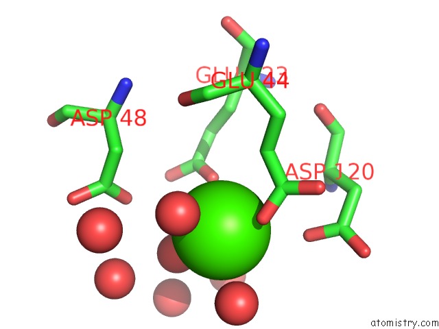
Mono view
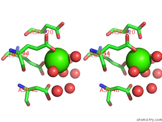
Stereo pair view
Mono view
Stereo pair view
A full contact list of Calcium with other atoms in the Ca binding
site number 1 of Crystal Structure of Nuclease Domain of Ribonuclase III From Mycobacterium Tuberculosis within 5.0Å range:
|
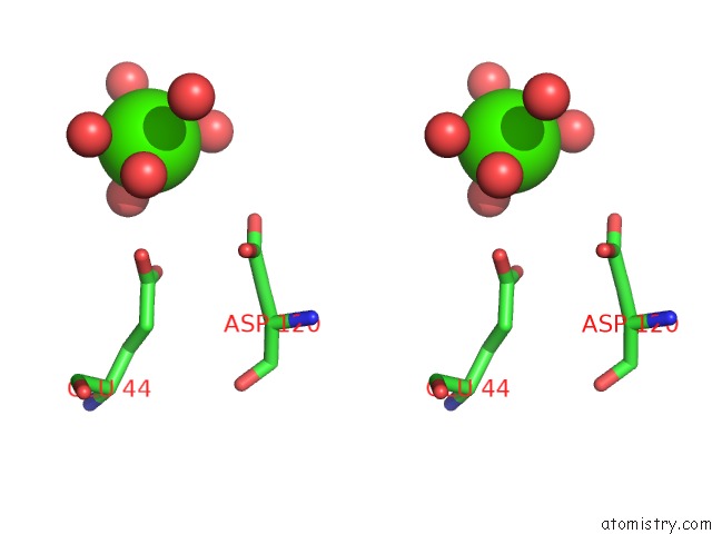
Calcium binding site 2 out of 2 in 2a11
Go back to
Calcium binding site 2 out
of 2 in the Crystal Structure of Nuclease Domain of Ribonuclase III From Mycobacterium Tuberculosis
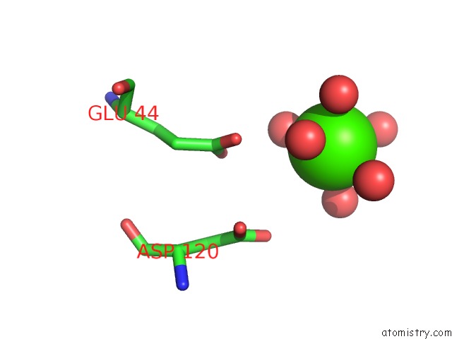
Mono view
Stereo pair view
Mono view
Stereo pair view
A full contact list of Calcium with other atoms in the Ca binding
site number 2 of Crystal Structure of Nuclease Domain of Ribonuclase III From Mycobacterium Tuberculosis within 5.0Å range:
|
Reference:
D.L.Akey,
J.M.Berger.
Structure of the Nuclease Domain of Ribonuclease III From M. Tuberculosis at 2.1 A Protein Sci. V. 14 2744 2005.
ISSN: ISSN 0961-8368
PubMed: 16155207
DOI: 10.1110/PS.051665905
Page generated: Fri Jul 12 08:30:40 2024
ISSN: ISSN 0961-8368
PubMed: 16155207
DOI: 10.1110/PS.051665905
Last articles
Zn in 9J0NZn in 9J0O
Zn in 9J0P
Zn in 9FJX
Zn in 9EKB
Zn in 9C0F
Zn in 9CAH
Zn in 9CH0
Zn in 9CH3
Zn in 9CH1