Calcium »
PDB 1zf0-2a2z »
2a2s »
Calcium in PDB 2a2s: Crystal Structure of Human Glutathione Transferase in Complex with S-Nitrosoglutathione in the Absence of Reducing Agent
Enzymatic activity of Crystal Structure of Human Glutathione Transferase in Complex with S-Nitrosoglutathione in the Absence of Reducing Agent
All present enzymatic activity of Crystal Structure of Human Glutathione Transferase in Complex with S-Nitrosoglutathione in the Absence of Reducing Agent:
2.5.1.18;
2.5.1.18;
Protein crystallography data
The structure of Crystal Structure of Human Glutathione Transferase in Complex with S-Nitrosoglutathione in the Absence of Reducing Agent, PDB code: 2a2s
was solved by
L.J.Parker,
C.J.Morton,
J.J.Adams,
M.W.Parker,
with X-Ray Crystallography technique. A brief refinement statistics is given in the table below:
| Resolution Low / High (Å) | 30.00 / 1.70 |
| Space group | C 1 2 1 |
| Cell size a, b, c (Å), α, β, γ (°) | 77.640, 89.640, 68.850, 90.00, 97.98, 90.00 |
| R / Rfree (%) | 17.8 / 20.8 |
Calcium Binding Sites:
The binding sites of Calcium atom in the Crystal Structure of Human Glutathione Transferase in Complex with S-Nitrosoglutathione in the Absence of Reducing Agent
(pdb code 2a2s). This binding sites where shown within
5.0 Angstroms radius around Calcium atom.
In total only one binding site of Calcium was determined in the Crystal Structure of Human Glutathione Transferase in Complex with S-Nitrosoglutathione in the Absence of Reducing Agent, PDB code: 2a2s:
In total only one binding site of Calcium was determined in the Crystal Structure of Human Glutathione Transferase in Complex with S-Nitrosoglutathione in the Absence of Reducing Agent, PDB code: 2a2s:
Calcium binding site 1 out of 1 in 2a2s
Go back to
Calcium binding site 1 out
of 1 in the Crystal Structure of Human Glutathione Transferase in Complex with S-Nitrosoglutathione in the Absence of Reducing Agent
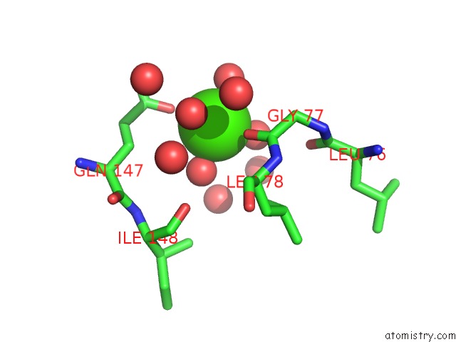
Mono view
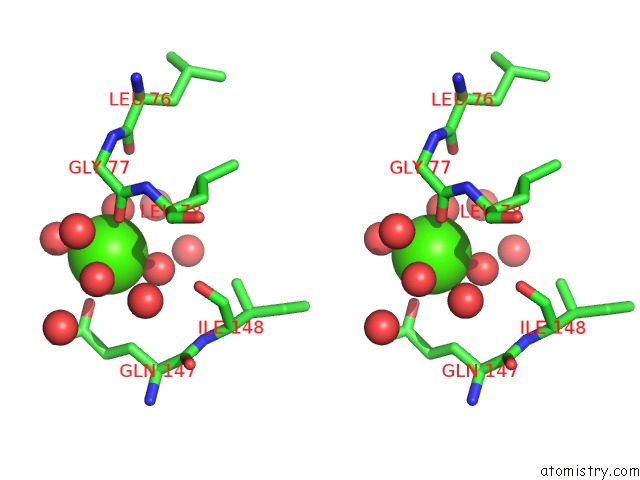
Stereo pair view
Mono view
Stereo pair view
A full contact list of Calcium with other atoms in the Ca binding
site number 1 of Crystal Structure of Human Glutathione Transferase in Complex with S-Nitrosoglutathione in the Absence of Reducing Agent within 5.0Å range:
|
Reference:
R.Tellez-Sanz,
E.Cesareo,
M.Nuccetelli,
A.M.Aguilera,
C.Baron,
L.J.Parker,
J.J.Adams,
C.J.Morton,
M.Lo Bello,
M.W.Parker,
L.Garcia-Fuentes.
Calorimetric and Structural Studies of the Nitric Oxide Carrier S-Nitrosoglutathione Bound to Human Glutathione Transferase P1-1 Protein Sci. V. 15 1093 2006.
ISSN: ISSN 0961-8368
PubMed: 16597834
DOI: 10.1110/PS.052055206
Page generated: Fri Jul 12 08:31:07 2024
ISSN: ISSN 0961-8368
PubMed: 16597834
DOI: 10.1110/PS.052055206
Last articles
Zn in 9J0NZn in 9J0O
Zn in 9J0P
Zn in 9FJX
Zn in 9EKB
Zn in 9C0F
Zn in 9CAH
Zn in 9CH0
Zn in 9CH3
Zn in 9CH1