Calcium »
PDB 1zf0-2a2z »
2a2z »
Calcium in PDB 2a2z: Crystal Structure of Human Deoxycytidine Kinase in Complex with Deoxycytidine and Uridine Diphosphate
Enzymatic activity of Crystal Structure of Human Deoxycytidine Kinase in Complex with Deoxycytidine and Uridine Diphosphate
All present enzymatic activity of Crystal Structure of Human Deoxycytidine Kinase in Complex with Deoxycytidine and Uridine Diphosphate:
2.7.1.74;
2.7.1.74;
Protein crystallography data
The structure of Crystal Structure of Human Deoxycytidine Kinase in Complex with Deoxycytidine and Uridine Diphosphate, PDB code: 2a2z
was solved by
M.H.Godsey,
S.Ort,
E.Sabini,
M.Konrad,
A.Lavie,
with X-Ray Crystallography technique. A brief refinement statistics is given in the table below:
| Resolution Low / High (Å) | 29.66 / 3.02 |
| Space group | P 21 21 21 |
| Cell size a, b, c (Å), α, β, γ (°) | 64.180, 110.850, 155.370, 90.00, 90.00, 90.00 |
| R / Rfree (%) | 24.6 / 30 |
Other elements in 2a2z:
The structure of Crystal Structure of Human Deoxycytidine Kinase in Complex with Deoxycytidine and Uridine Diphosphate also contains other interesting chemical elements:
| Magnesium | (Mg) | 4 atoms |
Calcium Binding Sites:
The binding sites of Calcium atom in the Crystal Structure of Human Deoxycytidine Kinase in Complex with Deoxycytidine and Uridine Diphosphate
(pdb code 2a2z). This binding sites where shown within
5.0 Angstroms radius around Calcium atom.
In total 2 binding sites of Calcium where determined in the Crystal Structure of Human Deoxycytidine Kinase in Complex with Deoxycytidine and Uridine Diphosphate, PDB code: 2a2z:
Jump to Calcium binding site number: 1; 2;
In total 2 binding sites of Calcium where determined in the Crystal Structure of Human Deoxycytidine Kinase in Complex with Deoxycytidine and Uridine Diphosphate, PDB code: 2a2z:
Jump to Calcium binding site number: 1; 2;
Calcium binding site 1 out of 2 in 2a2z
Go back to
Calcium binding site 1 out
of 2 in the Crystal Structure of Human Deoxycytidine Kinase in Complex with Deoxycytidine and Uridine Diphosphate
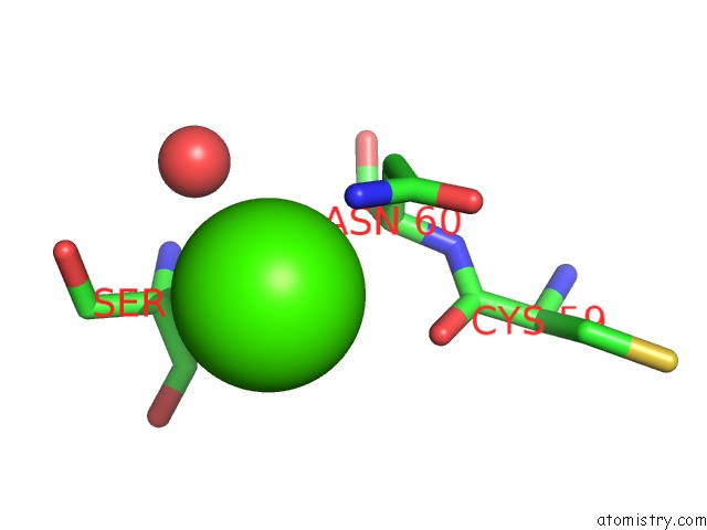
Mono view

Stereo pair view
Mono view
Stereo pair view
A full contact list of Calcium with other atoms in the Ca binding
site number 1 of Crystal Structure of Human Deoxycytidine Kinase in Complex with Deoxycytidine and Uridine Diphosphate within 5.0Å range:
|
Calcium binding site 2 out of 2 in 2a2z
Go back to
Calcium binding site 2 out
of 2 in the Crystal Structure of Human Deoxycytidine Kinase in Complex with Deoxycytidine and Uridine Diphosphate
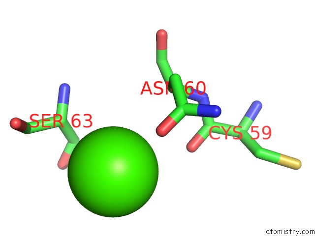
Mono view
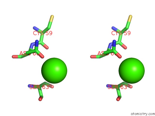
Stereo pair view
Mono view
Stereo pair view
A full contact list of Calcium with other atoms in the Ca binding
site number 2 of Crystal Structure of Human Deoxycytidine Kinase in Complex with Deoxycytidine and Uridine Diphosphate within 5.0Å range:
|
Reference:
M.H.Godsey,
S.Ort,
E.Sabini,
M.Konrad,
A.Lavie.
Structural Basis For the Preference of Utp Over Atp in Human Deoxycytidine Kinase: Illuminating the Role of Main-Chain Reorganization. Biochemistry V. 45 452 2006.
ISSN: ISSN 0006-2960
PubMed: 16401075
DOI: 10.1021/BI0518646
Page generated: Fri Jul 12 08:31:35 2024
ISSN: ISSN 0006-2960
PubMed: 16401075
DOI: 10.1021/BI0518646
Last articles
Zn in 9J0NZn in 9J0O
Zn in 9J0P
Zn in 9FJX
Zn in 9EKB
Zn in 9C0F
Zn in 9CAH
Zn in 9CH0
Zn in 9CH3
Zn in 9CH1