Calcium »
PDB 2a30-2agp »
2a4e »
Calcium in PDB 2a4e: Crystal Structure of Mouse Cadherin-11 EC1-2
Protein crystallography data
The structure of Crystal Structure of Mouse Cadherin-11 EC1-2, PDB code: 2a4e
was solved by
S.D.Patel,
C.Ciatto,
C.P.Chen,
F.Bahna,
N.Arkus,
M.Rajebhosale,
T.M.Jessell,
B.Honig,
S.R.Price,
L.Shapiro,
with X-Ray Crystallography technique. A brief refinement statistics is given in the table below:
| Resolution Low / High (Å) | 30.00 / 3.20 |
| Space group | P 62 2 2 |
| Cell size a, b, c (Å), α, β, γ (°) | 203.249, 203.249, 42.811, 90.00, 90.00, 120.00 |
| R / Rfree (%) | 21.1 / 24.7 |
Calcium Binding Sites:
The binding sites of Calcium atom in the Crystal Structure of Mouse Cadherin-11 EC1-2
(pdb code 2a4e). This binding sites where shown within
5.0 Angstroms radius around Calcium atom.
In total 3 binding sites of Calcium where determined in the Crystal Structure of Mouse Cadherin-11 EC1-2, PDB code: 2a4e:
Jump to Calcium binding site number: 1; 2; 3;
In total 3 binding sites of Calcium where determined in the Crystal Structure of Mouse Cadherin-11 EC1-2, PDB code: 2a4e:
Jump to Calcium binding site number: 1; 2; 3;
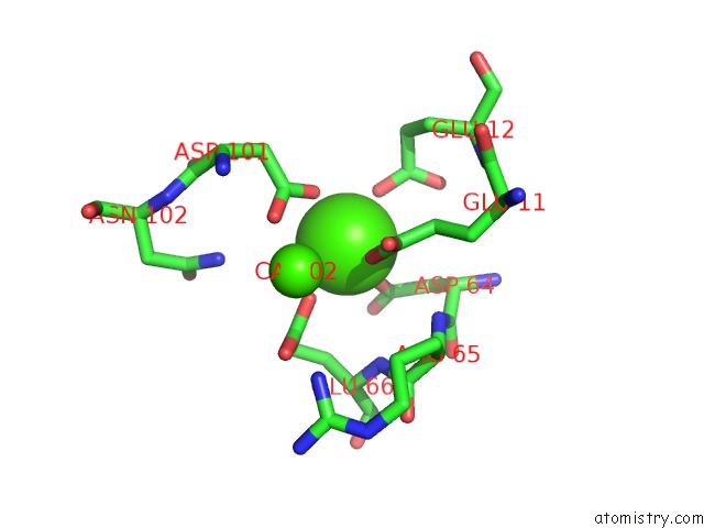
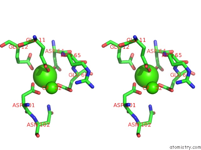
Calcium binding site 1 out of 3 in 2a4e
Go back to
Calcium binding site 1 out
of 3 in the Crystal Structure of Mouse Cadherin-11 EC1-2
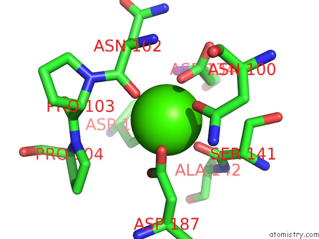
Mono view
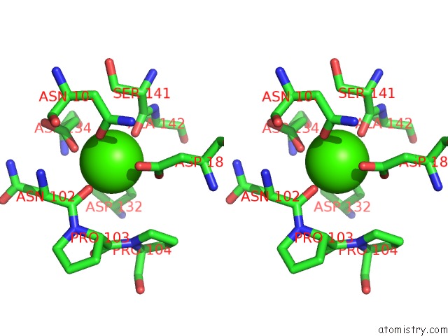
Stereo pair view
Mono view
Stereo pair view
A full contact list of Calcium with other atoms in the Ca binding
site number 1 of Crystal Structure of Mouse Cadherin-11 EC1-2 within 5.0Å range:
|
Calcium binding site 2 out of 3 in 2a4e
Go back to
Calcium binding site 2 out
of 3 in the Crystal Structure of Mouse Cadherin-11 EC1-2
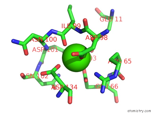
Mono view
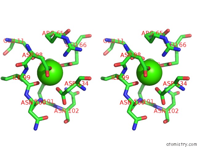
Stereo pair view
Mono view
Stereo pair view
A full contact list of Calcium with other atoms in the Ca binding
site number 2 of Crystal Structure of Mouse Cadherin-11 EC1-2 within 5.0Å range:
|
Calcium binding site 3 out of 3 in 2a4e
Go back to
Calcium binding site 3 out
of 3 in the Crystal Structure of Mouse Cadherin-11 EC1-2
Mono view
Stereo pair view
Mono view
Stereo pair view
A full contact list of Calcium with other atoms in the Ca binding
site number 3 of Crystal Structure of Mouse Cadherin-11 EC1-2 within 5.0Å range:
|
Reference:
S.D.Patel,
C.Ciatto,
C.P.Chen,
F.Bahna,
M.Rajebhosale,
N.Arkus,
I.Schieren,
T.M.Jessell,
B.Honig,
S.R.Price,
L.Shapiro.
Type II Cadherin Ectodomain Structures: Implications For Classical Cadherin Specificity. Cell(Cambridge,Mass.) V. 124 1255 2006.
ISSN: ISSN 0092-8674
PubMed: 16564015
DOI: 10.1016/J.CELL.2005.12.046
Page generated: Fri Jul 12 08:34:51 2024
ISSN: ISSN 0092-8674
PubMed: 16564015
DOI: 10.1016/J.CELL.2005.12.046
Last articles
Zn in 9MJ5Zn in 9HNW
Zn in 9G0L
Zn in 9FNE
Zn in 9DZN
Zn in 9E0I
Zn in 9D32
Zn in 9DAK
Zn in 8ZXC
Zn in 8ZUF