Calcium in PDB 2a62: Crystal Structure of Mouse Cadherin-8 EC1-3
Protein crystallography data
The structure of Crystal Structure of Mouse Cadherin-8 EC1-3, PDB code: 2a62
was solved by
S.D.Patel,
C.Ciatto,
C.P.Chen,
F.Bahna,
N.Arkus,
I.Schieren,
T.M.Jessell,
B.Honig,
S.R.Price,
L.Shapiro,
with X-Ray Crystallography technique. A brief refinement statistics is given in the table below:
Resolution Low / High (Å)
|
20.00 /
4.50
|
Space group
|
P 41 2 2
|
Cell size a, b, c (Å), α, β, γ (°)
|
75.818,
75.818,
233.760,
90.00,
90.00,
90.00
|
R / Rfree (%)
|
27.1 /
34.6
|
Calcium Binding Sites:
The binding sites of Calcium atom in the Crystal Structure of Mouse Cadherin-8 EC1-3
(pdb code 2a62). This binding sites where shown within
5.0 Angstroms radius around Calcium atom.
In total 6 binding sites of Calcium where determined in the
Crystal Structure of Mouse Cadherin-8 EC1-3, PDB code: 2a62:
Jump to Calcium binding site number:
1;
2;
3;
4;
5;
6;
Calcium binding site 1 out
of 6 in 2a62
Go back to
Calcium Binding Sites List in 2a62
Calcium binding site 1 out
of 6 in the Crystal Structure of Mouse Cadherin-8 EC1-3
 Mono view
 Stereo pair view
|
A full contact list of Calcium with other atoms in the Ca binding
site number 1 of Crystal Structure of Mouse Cadherin-8 EC1-3 within 5.0Å range:
probe
|
atom
|
residue
|
distance (Å)
|
B
|
Occ
|
A:Ca323
b:0.6
occ:1.00
|
OD2
|
A:ASP132
|
1.9
|
0.2
|
1.0
|
O
|
A:ASN102
|
2.2
|
0.2
|
1.0
|
OD1
|
A:ASN100
|
2.2
|
0.3
|
1.0
|
OD1
|
A:ASP132
|
2.5
|
0.8
|
1.0
|
CG
|
A:ASP132
|
2.5
|
0.8
|
1.0
|
OD2
|
A:ASP187
|
2.9
|
0.9
|
1.0
|
OD2
|
A:ASP134
|
3.0
|
0.3
|
1.0
|
O
|
A:SER141
|
3.1
|
1.0
|
1.0
|
C
|
A:ASN102
|
3.2
|
0.3
|
1.0
|
CB
|
A:ASP134
|
3.3
|
0.5
|
1.0
|
CG
|
A:ASN100
|
3.3
|
0.3
|
1.0
|
OD1
|
A:ASP187
|
3.4
|
1.0
|
1.0
|
CG
|
A:ASP187
|
3.5
|
0.9
|
1.0
|
CG
|
A:ASP134
|
3.5
|
0.3
|
1.0
|
CB
|
A:ASN102
|
3.8
|
0.5
|
1.0
|
CA
|
A:ASN102
|
3.8
|
0.4
|
1.0
|
N
|
A:ASN102
|
3.9
|
0.4
|
1.0
|
N
|
A:ASP134
|
4.0
|
0.3
|
1.0
|
ND2
|
A:ASN100
|
4.0
|
0.5
|
1.0
|
CB
|
A:ASP132
|
4.0
|
0.5
|
1.0
|
N
|
A:ALA103
|
4.2
|
0.4
|
1.0
|
CA
|
A:ASP134
|
4.2
|
0.6
|
1.0
|
C
|
A:SER141
|
4.3
|
0.1
|
1.0
|
CD2
|
A:LEU195
|
4.3
|
0.6
|
1.0
|
CA
|
A:ASN100
|
4.4
|
0.4
|
1.0
|
CB
|
A:ASN100
|
4.4
|
0.3
|
1.0
|
N
|
A:ALA133
|
4.5
|
0.9
|
1.0
|
C
|
A:ASN100
|
4.6
|
0.3
|
1.0
|
OD1
|
A:ASP134
|
4.7
|
0.7
|
1.0
|
CA
|
A:ALA103
|
4.7
|
0.6
|
1.0
|
CA
|
A:ASP132
|
4.8
|
0.8
|
1.0
|
CB
|
A:ASP187
|
4.8
|
0.9
|
1.0
|
C
|
A:ALA103
|
4.9
|
0.6
|
1.0
|
O
|
A:ASN100
|
4.9
|
0.3
|
1.0
|
ND2
|
A:ASN102
|
4.9
|
0.8
|
1.0
|
CG
|
A:ASN102
|
4.9
|
0.8
|
1.0
|
O
|
A:ALA103
|
4.9
|
0.9
|
1.0
|
C
|
A:ASP132
|
4.9
|
0.9
|
1.0
|
|
Calcium binding site 2 out
of 6 in 2a62
Go back to
Calcium Binding Sites List in 2a62
Calcium binding site 2 out
of 6 in the Crystal Structure of Mouse Cadherin-8 EC1-3
 Mono view
 Stereo pair view
|
A full contact list of Calcium with other atoms in the Ca binding
site number 2 of Crystal Structure of Mouse Cadherin-8 EC1-3 within 5.0Å range:
probe
|
atom
|
residue
|
distance (Å)
|
B
|
Occ
|
A:Ca324
b:0.2
occ:1.00
|
OD1
|
A:ASP101
|
2.0
|
0.4
|
1.0
|
OD2
|
A:ASP98
|
2.0
|
0.1
|
1.0
|
ND2
|
A:ASN102
|
2.3
|
0.8
|
1.0
|
OE2
|
A:GLU11
|
2.3
|
0.8
|
1.0
|
O
|
A:ILE99
|
2.3
|
0.7
|
1.0
|
OE2
|
A:GLU66
|
2.8
|
0.6
|
1.0
|
CG
|
A:ASP101
|
3.2
|
0.8
|
1.0
|
CG
|
A:ASP98
|
3.2
|
0.9
|
1.0
|
CD
|
A:GLU11
|
3.3
|
0.9
|
1.0
|
OD1
|
A:ASP134
|
3.3
|
0.7
|
1.0
|
OD2
|
A:ASP134
|
3.3
|
0.3
|
1.0
|
CA
|
A:CA325
|
3.4
|
0.1
|
1.0
|
CG
|
A:ASN102
|
3.4
|
0.8
|
1.0
|
CD
|
A:GLU66
|
3.4
|
0.6
|
1.0
|
C
|
A:ILE99
|
3.5
|
0.6
|
1.0
|
CG
|
A:ASP134
|
3.6
|
0.3
|
1.0
|
N
|
A:ILE99
|
3.7
|
0.6
|
1.0
|
OD2
|
A:ASP101
|
3.7
|
0.8
|
1.0
|
OE1
|
A:GLU66
|
3.8
|
0.4
|
1.0
|
OE1
|
A:GLU11
|
3.8
|
0.2
|
1.0
|
OD1
|
A:ASP98
|
3.9
|
0.9
|
1.0
|
OD1
|
A:ASN102
|
3.9
|
0.2
|
1.0
|
N
|
A:ASP101
|
4.0
|
0.3
|
1.0
|
CA
|
A:ILE99
|
4.2
|
0.6
|
1.0
|
CG
|
A:GLU11
|
4.3
|
0.9
|
1.0
|
CB
|
A:ASP101
|
4.3
|
0.1
|
1.0
|
CA
|
A:ASP98
|
4.4
|
1.0
|
1.0
|
CB
|
A:ASP98
|
4.4
|
0.0
|
1.0
|
C
|
A:ASP98
|
4.4
|
0.7
|
1.0
|
N
|
A:ASN102
|
4.4
|
0.4
|
1.0
|
CG
|
A:GLU66
|
4.5
|
0.8
|
1.0
|
CG2
|
A:ILE99
|
4.5
|
0.7
|
1.0
|
N
|
A:ASN100
|
4.6
|
0.5
|
1.0
|
CB
|
A:ASN102
|
4.6
|
0.5
|
1.0
|
CA
|
A:ASP101
|
4.6
|
0.3
|
1.0
|
CA
|
A:ASN100
|
4.9
|
0.4
|
1.0
|
C
|
A:ASP101
|
4.9
|
0.4
|
1.0
|
CB
|
A:ASP134
|
4.9
|
0.5
|
1.0
|
C
|
A:ASN100
|
4.9
|
0.3
|
1.0
|
|
Calcium binding site 3 out
of 6 in 2a62
Go back to
Calcium Binding Sites List in 2a62
Calcium binding site 3 out
of 6 in the Crystal Structure of Mouse Cadherin-8 EC1-3
 Mono view
 Stereo pair view
|
A full contact list of Calcium with other atoms in the Ca binding
site number 3 of Crystal Structure of Mouse Cadherin-8 EC1-3 within 5.0Å range:
probe
|
atom
|
residue
|
distance (Å)
|
B
|
Occ
|
A:Ca325
b:0.1
occ:1.00
|
OE1
|
A:GLU66
|
2.1
|
0.4
|
1.0
|
OE2
|
A:GLU12
|
2.4
|
0.1
|
1.0
|
OD2
|
A:ASP64
|
2.4
|
0.2
|
1.0
|
OE2
|
A:GLU11
|
2.6
|
0.8
|
1.0
|
OD2
|
A:ASP101
|
2.6
|
0.8
|
1.0
|
OE2
|
A:GLU66
|
2.8
|
0.6
|
1.0
|
CD
|
A:GLU66
|
2.8
|
0.6
|
1.0
|
OE1
|
A:GLU11
|
2.9
|
0.2
|
1.0
|
CD
|
A:GLU11
|
3.1
|
0.9
|
1.0
|
OD1
|
A:ASP101
|
3.3
|
0.4
|
1.0
|
CD
|
A:GLU12
|
3.3
|
0.2
|
1.0
|
CG
|
A:ASP64
|
3.3
|
0.9
|
1.0
|
CG
|
A:ASP101
|
3.3
|
0.8
|
1.0
|
CA
|
A:CA324
|
3.4
|
0.2
|
1.0
|
CG
|
A:GLU12
|
3.6
|
0.1
|
1.0
|
OD1
|
A:ASP64
|
3.9
|
0.9
|
1.0
|
OD1
|
A:ASN102
|
4.1
|
0.2
|
1.0
|
CA
|
A:ASP64
|
4.2
|
0.4
|
1.0
|
CB
|
A:ASP64
|
4.2
|
0.3
|
1.0
|
N
|
A:ARG65
|
4.3
|
0.8
|
1.0
|
CG
|
A:GLU66
|
4.3
|
0.8
|
1.0
|
ND2
|
A:ASN102
|
4.3
|
0.8
|
1.0
|
OE1
|
A:GLU12
|
4.4
|
0.5
|
1.0
|
CG
|
A:GLU11
|
4.5
|
0.9
|
1.0
|
CG
|
A:ASN102
|
4.6
|
0.8
|
1.0
|
C
|
A:ASP64
|
4.7
|
0.5
|
1.0
|
CB
|
A:ASP101
|
4.8
|
0.1
|
1.0
|
N
|
A:GLU66
|
4.8
|
0.7
|
1.0
|
OD2
|
A:ASP98
|
4.8
|
0.1
|
1.0
|
O
|
A:LEU63
|
4.9
|
0.4
|
1.0
|
CB
|
A:GLU12
|
4.9
|
0.3
|
1.0
|
CB
|
A:GLU66
|
5.0
|
0.9
|
1.0
|
|
Calcium binding site 4 out
of 6 in 2a62
Go back to
Calcium Binding Sites List in 2a62
Calcium binding site 4 out
of 6 in the Crystal Structure of Mouse Cadherin-8 EC1-3
 Mono view
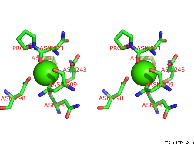
 Stereo pair view
|
A full contact list of Calcium with other atoms in the Ca binding
site number 4 of Crystal Structure of Mouse Cadherin-8 EC1-3 within 5.0Å range:
probe
|
atom
|
residue
|
distance (Å)
|
B
|
Occ
|
A:Ca326
b:0.5
occ:1.00
|
OD2
|
A:ASP241
|
1.8
|
0.1
|
1.0
|
OD1
|
A:ASN209
|
1.9
|
0.7
|
1.0
|
O
|
A:ASN211
|
2.2
|
0.5
|
1.0
|
OD1
|
A:ASP241
|
2.2
|
0.7
|
1.0
|
CG
|
A:ASP241
|
2.3
|
0.7
|
1.0
|
OD1
|
A:ASN298
|
2.5
|
0.0
|
1.0
|
O
|
A:ASN247
|
2.9
|
0.8
|
1.0
|
CG
|
A:ASN209
|
2.9
|
0.0
|
1.0
|
C
|
A:ASN211
|
3.3
|
0.4
|
1.0
|
CG
|
A:ASN298
|
3.4
|
0.1
|
1.0
|
CB
|
A:ASP243
|
3.5
|
0.8
|
1.0
|
ND2
|
A:ASN209
|
3.6
|
0.0
|
1.0
|
CB
|
A:ASN211
|
3.7
|
0.5
|
1.0
|
O
|
A:ASN209
|
3.7
|
0.3
|
1.0
|
CB
|
A:ASP241
|
3.8
|
1.0
|
1.0
|
CA
|
A:ASN211
|
3.8
|
0.5
|
1.0
|
OD2
|
A:ASP243
|
3.9
|
0.9
|
1.0
|
N
|
A:ASN211
|
3.9
|
0.5
|
1.0
|
ND2
|
A:ASN298
|
4.0
|
0.5
|
1.0
|
CG
|
A:ASP243
|
4.0
|
0.4
|
1.0
|
CB
|
A:ASN209
|
4.1
|
0.1
|
1.0
|
C
|
A:ASN247
|
4.1
|
0.8
|
1.0
|
C
|
A:ASN209
|
4.1
|
0.1
|
1.0
|
CA
|
A:ASN209
|
4.2
|
0.1
|
1.0
|
N
|
A:PRO212
|
4.4
|
0.5
|
1.0
|
O
|
A:ASP243
|
4.4
|
0.2
|
1.0
|
CB
|
A:ASN298
|
4.5
|
0.9
|
1.0
|
C
|
A:PRO212
|
4.6
|
0.6
|
1.0
|
CA
|
A:PRO212
|
4.7
|
0.6
|
1.0
|
CA
|
A:ASP243
|
4.7
|
1.0
|
1.0
|
O
|
A:PRO212
|
4.7
|
0.5
|
1.0
|
CB
|
A:ASN247
|
4.7
|
0.7
|
1.0
|
CA
|
A:ASP241
|
4.7
|
0.0
|
1.0
|
N
|
A:ASP243
|
4.8
|
0.0
|
1.0
|
CA
|
A:ASN247
|
4.9
|
0.7
|
1.0
|
|
Calcium binding site 5 out
of 6 in 2a62
Go back to
Calcium Binding Sites List in 2a62
Calcium binding site 5 out
of 6 in the Crystal Structure of Mouse Cadherin-8 EC1-3
 Mono view
 Stereo pair view
|
A full contact list of Calcium with other atoms in the Ca binding
site number 5 of Crystal Structure of Mouse Cadherin-8 EC1-3 within 5.0Å range:
probe
|
atom
|
residue
|
distance (Å)
|
B
|
Occ
|
A:Ca327
b:0.6
occ:1.00
|
CA
|
A:CA328
|
1.8
|
0.9
|
1.0
|
OD1
|
A:ASP210
|
2.1
|
0.5
|
1.0
|
O
|
A:ASP207
|
2.1
|
0.5
|
1.0
|
OD2
|
A:ASP207
|
2.2
|
0.7
|
1.0
|
O
|
A:VAL208
|
2.4
|
0.4
|
1.0
|
OD2
|
A:ASP243
|
2.9
|
0.9
|
1.0
|
OE1
|
A:GLU117
|
3.0
|
0.8
|
1.0
|
C
|
A:ASP207
|
3.1
|
0.5
|
1.0
|
CG
|
A:ASP243
|
3.1
|
0.4
|
1.0
|
C
|
A:VAL208
|
3.2
|
0.2
|
1.0
|
CG
|
A:ASP210
|
3.3
|
0.1
|
1.0
|
CG
|
A:ASP207
|
3.3
|
0.7
|
1.0
|
OD1
|
A:ASP243
|
3.4
|
0.1
|
1.0
|
CG1
|
A:VAL208
|
3.5
|
0.0
|
1.0
|
N
|
A:ASP210
|
3.5
|
0.9
|
1.0
|
CD
|
A:GLU117
|
3.7
|
0.1
|
1.0
|
CA
|
A:VAL208
|
3.8
|
0.1
|
1.0
|
OE2
|
A:GLU117
|
3.8
|
0.8
|
1.0
|
N
|
A:VAL208
|
3.8
|
0.8
|
1.0
|
CB
|
A:ASP243
|
3.9
|
0.8
|
1.0
|
CB
|
A:VAL208
|
3.9
|
0.2
|
1.0
|
CA
|
A:ASP207
|
4.0
|
0.4
|
1.0
|
OD2
|
A:ASP210
|
4.1
|
0.2
|
1.0
|
NH2
|
A:ARG173
|
4.1
|
0.5
|
1.0
|
N
|
A:ASN209
|
4.1
|
0.2
|
1.0
|
OD1
|
A:ASP207
|
4.1
|
0.4
|
1.0
|
CB
|
A:ASP210
|
4.2
|
0.6
|
1.0
|
CB
|
A:ASP207
|
4.2
|
0.6
|
1.0
|
OE1
|
A:GLU174
|
4.3
|
0.8
|
1.0
|
OE2
|
A:GLU174
|
4.3
|
0.3
|
1.0
|
CA
|
A:ASP210
|
4.3
|
0.7
|
1.0
|
CA
|
A:ASN209
|
4.3
|
0.1
|
1.0
|
C
|
A:ASN209
|
4.3
|
0.1
|
1.0
|
CD
|
A:GLU174
|
4.4
|
0.8
|
1.0
|
CA
|
A:ASP243
|
4.5
|
1.0
|
1.0
|
C
|
A:ASP210
|
4.7
|
0.6
|
1.0
|
CZ
|
A:ARG173
|
4.9
|
0.5
|
1.0
|
|
Calcium binding site 6 out
of 6 in 2a62
Go back to
Calcium Binding Sites List in 2a62
Calcium binding site 6 out
of 6 in the Crystal Structure of Mouse Cadherin-8 EC1-3
 Mono view
 Stereo pair view
|
A full contact list of Calcium with other atoms in the Ca binding
site number 6 of Crystal Structure of Mouse Cadherin-8 EC1-3 within 5.0Å range:
probe
|
atom
|
residue
|
distance (Å)
|
B
|
Occ
|
A:Ca328
b:0.9
occ:1.00
|
CA
|
A:CA327
|
1.8
|
0.6
|
1.0
|
OE1
|
A:GLU117
|
2.3
|
0.8
|
1.0
|
OD2
|
A:ASP207
|
2.5
|
0.7
|
1.0
|
OD1
|
A:ASP210
|
2.6
|
0.5
|
1.0
|
NH2
|
A:ARG173
|
2.7
|
0.5
|
1.0
|
OE2
|
A:GLU174
|
2.7
|
0.3
|
1.0
|
CD
|
A:GLU174
|
3.0
|
0.8
|
1.0
|
OD1
|
A:ASP243
|
3.0
|
0.1
|
1.0
|
CD
|
A:GLU117
|
3.2
|
0.1
|
1.0
|
OE1
|
A:GLU174
|
3.2
|
0.8
|
1.0
|
O
|
A:ASP207
|
3.3
|
0.5
|
1.0
|
CG
|
A:ASP243
|
3.3
|
0.4
|
1.0
|
OE2
|
A:GLU117
|
3.4
|
0.8
|
1.0
|
CZ
|
A:ARG173
|
3.5
|
0.5
|
1.0
|
OD2
|
A:ASP243
|
3.6
|
0.9
|
1.0
|
CG
|
A:ASP210
|
3.7
|
0.1
|
1.0
|
CG
|
A:ASP207
|
3.7
|
0.7
|
1.0
|
NE
|
A:ARG173
|
3.7
|
0.5
|
1.0
|
CG
|
A:GLU174
|
3.8
|
0.1
|
1.0
|
O
|
A:VAL208
|
4.0
|
0.4
|
1.0
|
OD2
|
A:ASP210
|
4.0
|
0.2
|
1.0
|
CB
|
A:ASP243
|
4.1
|
0.8
|
1.0
|
C
|
A:ASP207
|
4.2
|
0.5
|
1.0
|
CA
|
A:ASP243
|
4.3
|
1.0
|
1.0
|
OD1
|
A:ASP207
|
4.4
|
0.4
|
1.0
|
OD2
|
A:ASP172
|
4.5
|
0.2
|
1.0
|
CA
|
A:ASP207
|
4.6
|
0.4
|
1.0
|
NH1
|
A:ARG173
|
4.6
|
0.6
|
1.0
|
CG
|
A:GLU117
|
4.6
|
0.8
|
1.0
|
CB
|
A:ARG173
|
4.8
|
0.8
|
1.0
|
CB
|
A:ASP207
|
4.8
|
0.6
|
1.0
|
N
|
A:GLU174
|
4.8
|
0.1
|
1.0
|
N
|
A:ASP210
|
4.9
|
0.9
|
1.0
|
C
|
A:VAL208
|
4.9
|
0.2
|
1.0
|
CB
|
A:ASP210
|
5.0
|
0.6
|
1.0
|
|
Reference:
S.D.Patel,
C.Ciatto,
C.P.Chen,
F.Bahna,
M.Rajebhosale,
N.Arkus,
I.Schieren,
T.M.Jessell,
B.Honig,
S.R.Price,
L.Shapiro.
Type II Cadherin Ectodomain Structures: Implications For Classical Cadherin Specificity. Cell(Cambridge,Mass.) V. 124 1255 2006.
ISSN: ISSN 0092-8674
PubMed: 16564015
DOI: 10.1016/J.CELL.2005.12.046
Page generated: Fri Jul 12 08:35:34 2024
|