Calcium »
PDB 2a30-2agp »
2afb »
Calcium in PDB 2afb: Crystal Structure of 2-Dehydro-3- Deoxygluconokinase (Ec 2.7.1.45) (TM0067) From Thermotoga Maritima at 2.05 A Resolution
Protein crystallography data
The structure of Crystal Structure of 2-Dehydro-3- Deoxygluconokinase (Ec 2.7.1.45) (TM0067) From Thermotoga Maritima at 2.05 A Resolution, PDB code: 2afb
was solved by
Joint Center For Structural Genomics (Jcsg),
with X-Ray Crystallography technique. A brief refinement statistics is given in the table below:
| Resolution Low / High (Å) | 48.63 / 2.05 |
| Space group | H 3 2 |
| Cell size a, b, c (Å), α, β, γ (°) | 120.993, 120.993, 260.300, 90.00, 90.00, 120.00 |
| R / Rfree (%) | 17.6 / 23 |
Other elements in 2afb:
The structure of Crystal Structure of 2-Dehydro-3- Deoxygluconokinase (Ec 2.7.1.45) (TM0067) From Thermotoga Maritima at 2.05 A Resolution also contains other interesting chemical elements:
| Nickel | (Ni) | 6 atoms |
Calcium Binding Sites:
The binding sites of Calcium atom in the Crystal Structure of 2-Dehydro-3- Deoxygluconokinase (Ec 2.7.1.45) (TM0067) From Thermotoga Maritima at 2.05 A Resolution
(pdb code 2afb). This binding sites where shown within
5.0 Angstroms radius around Calcium atom.
In total 4 binding sites of Calcium where determined in the Crystal Structure of 2-Dehydro-3- Deoxygluconokinase (Ec 2.7.1.45) (TM0067) From Thermotoga Maritima at 2.05 A Resolution, PDB code: 2afb:
Jump to Calcium binding site number: 1; 2; 3; 4;
In total 4 binding sites of Calcium where determined in the Crystal Structure of 2-Dehydro-3- Deoxygluconokinase (Ec 2.7.1.45) (TM0067) From Thermotoga Maritima at 2.05 A Resolution, PDB code: 2afb:
Jump to Calcium binding site number: 1; 2; 3; 4;
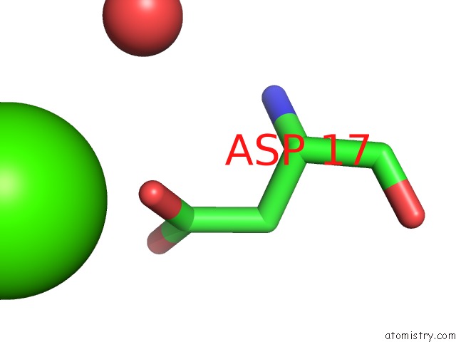
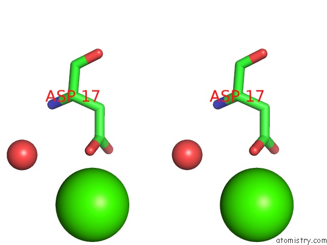
Calcium binding site 1 out of 4 in 2afb
Go back to
Calcium binding site 1 out
of 4 in the Crystal Structure of 2-Dehydro-3- Deoxygluconokinase (Ec 2.7.1.45) (TM0067) From Thermotoga Maritima at 2.05 A Resolution
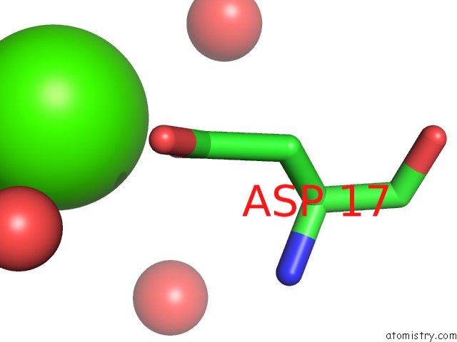
Mono view
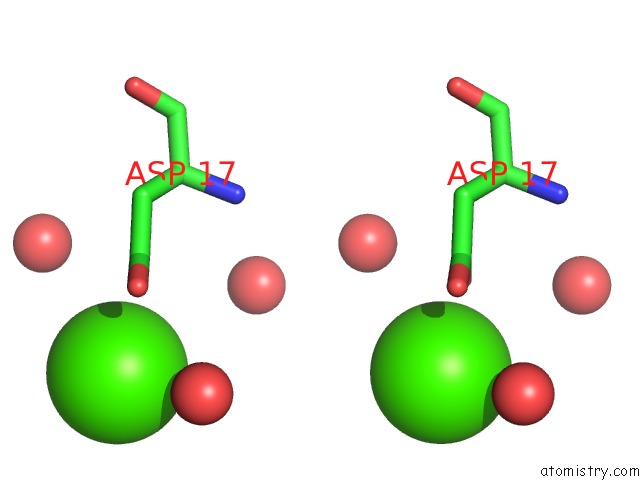
Stereo pair view
Mono view
Stereo pair view
A full contact list of Calcium with other atoms in the Ca binding
site number 1 of Crystal Structure of 2-Dehydro-3- Deoxygluconokinase (Ec 2.7.1.45) (TM0067) From Thermotoga Maritima at 2.05 A Resolution within 5.0Å range:
|
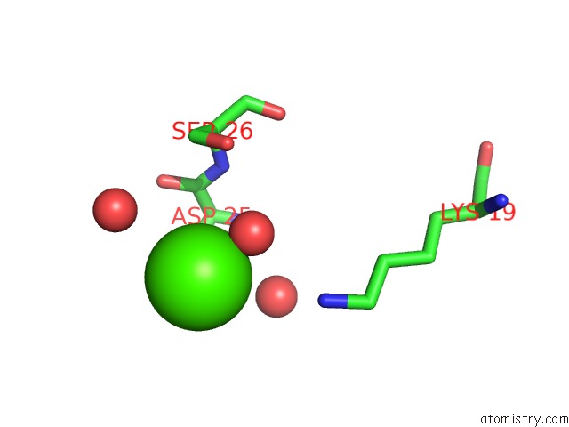
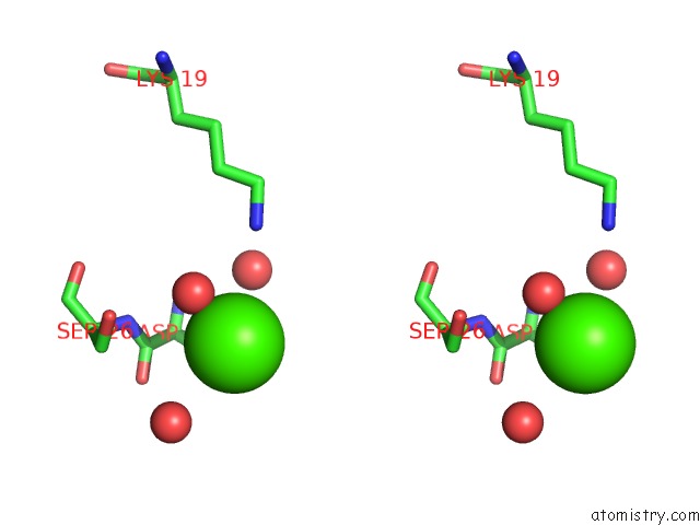
Calcium binding site 2 out of 4 in 2afb
Go back to
Calcium binding site 2 out
of 4 in the Crystal Structure of 2-Dehydro-3- Deoxygluconokinase (Ec 2.7.1.45) (TM0067) From Thermotoga Maritima at 2.05 A Resolution
Mono view
Stereo pair view
Mono view
Stereo pair view
A full contact list of Calcium with other atoms in the Ca binding
site number 2 of Crystal Structure of 2-Dehydro-3- Deoxygluconokinase (Ec 2.7.1.45) (TM0067) From Thermotoga Maritima at 2.05 A Resolution within 5.0Å range:
|
Calcium binding site 3 out of 4 in 2afb
Go back to
Calcium binding site 3 out
of 4 in the Crystal Structure of 2-Dehydro-3- Deoxygluconokinase (Ec 2.7.1.45) (TM0067) From Thermotoga Maritima at 2.05 A Resolution
Mono view
Stereo pair view
Mono view
Stereo pair view
A full contact list of Calcium with other atoms in the Ca binding
site number 3 of Crystal Structure of 2-Dehydro-3- Deoxygluconokinase (Ec 2.7.1.45) (TM0067) From Thermotoga Maritima at 2.05 A Resolution within 5.0Å range:
|
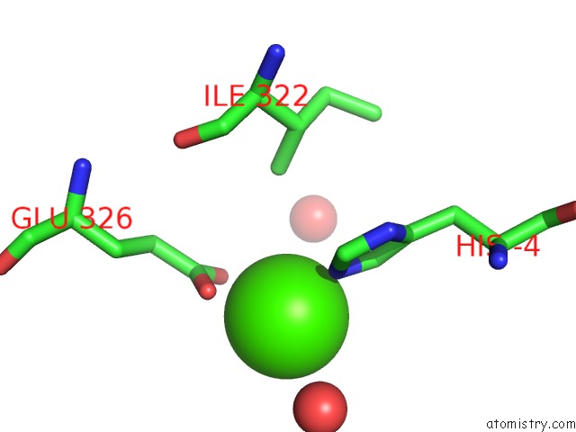
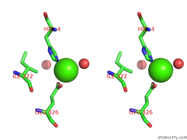
Calcium binding site 4 out of 4 in 2afb
Go back to
Calcium binding site 4 out
of 4 in the Crystal Structure of 2-Dehydro-3- Deoxygluconokinase (Ec 2.7.1.45) (TM0067) From Thermotoga Maritima at 2.05 A Resolution
Mono view
Stereo pair view
Mono view
Stereo pair view
A full contact list of Calcium with other atoms in the Ca binding
site number 4 of Crystal Structure of 2-Dehydro-3- Deoxygluconokinase (Ec 2.7.1.45) (TM0067) From Thermotoga Maritima at 2.05 A Resolution within 5.0Å range:
|
Reference:
I.I.Mathews,
D.Mcmullan,
M.D.Miller,
J.M.Canaves,
M.A.Elsliger,
R.Floyd,
S.K.Grzechnik,
L.Jaroszewski,
H.E.Klock,
E.Koesema,
J.S.Kovarik,
A.Kreusch,
P.Kuhn,
T.M.Mcphillips,
A.T.Morse,
K.Quijano,
C.L.Rife,
R.Schwarzenbacher,
G.Spraggon,
R.C.Stevens,
H.Van Den Bedem,
D.Weekes,
G.Wolf,
K.O.Hodgson,
J.Wooley,
A.M.Deacon,
A.Godzik,
S.A.Lesley,
I.A.Wilson.
Crystal Structure of 2-Keto-3-Deoxygluconate Kinase (TM0067) From Thermotoga Maritima at 2.05 A Resolution. Proteins V. 70 603 2007.
ISSN: ISSN 0887-3585
PubMed: 18004772
DOI: 10.1002/PROT.21842
Page generated: Fri Jul 12 08:41:22 2024
ISSN: ISSN 0887-3585
PubMed: 18004772
DOI: 10.1002/PROT.21842
Last articles
Zn in 9J0NZn in 9J0O
Zn in 9J0P
Zn in 9FJX
Zn in 9EKB
Zn in 9C0F
Zn in 9CAH
Zn in 9CH0
Zn in 9CH3
Zn in 9CH1