Calcium »
PDB 2a30-2agp »
2agi »
Calcium in PDB 2agi: The Leupeptin-Trypsin Covalent Complex at 1.14 A Resolution
Enzymatic activity of The Leupeptin-Trypsin Covalent Complex at 1.14 A Resolution
All present enzymatic activity of The Leupeptin-Trypsin Covalent Complex at 1.14 A Resolution:
3.4.21.4;
3.4.21.4;
Protein crystallography data
The structure of The Leupeptin-Trypsin Covalent Complex at 1.14 A Resolution, PDB code: 2agi
was solved by
E.S.Radisky,
J.M.Lee,
C.J.Lu,
D.E.Koshland Jr.,
with X-Ray Crystallography technique. A brief refinement statistics is given in the table below:
| Resolution Low / High (Å) | 46.63 / 1.14 |
| Space group | P 21 21 21 |
| Cell size a, b, c (Å), α, β, γ (°) | 61.888, 63.562, 69.102, 90.00, 90.00, 90.00 |
| R / Rfree (%) | 11.6 / 13.8 |
Calcium Binding Sites:
The binding sites of Calcium atom in the The Leupeptin-Trypsin Covalent Complex at 1.14 A Resolution
(pdb code 2agi). This binding sites where shown within
5.0 Angstroms radius around Calcium atom.
In total only one binding site of Calcium was determined in the The Leupeptin-Trypsin Covalent Complex at 1.14 A Resolution, PDB code: 2agi:
In total only one binding site of Calcium was determined in the The Leupeptin-Trypsin Covalent Complex at 1.14 A Resolution, PDB code: 2agi:
Calcium binding site 1 out of 1 in 2agi
Go back to
Calcium binding site 1 out
of 1 in the The Leupeptin-Trypsin Covalent Complex at 1.14 A Resolution
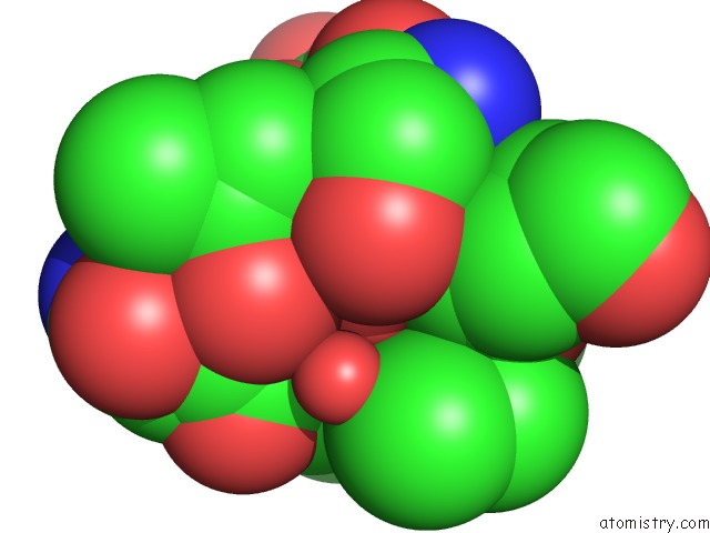
Mono view
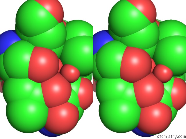
Stereo pair view
Mono view
Stereo pair view
A full contact list of Calcium with other atoms in the Ca binding
site number 1 of The Leupeptin-Trypsin Covalent Complex at 1.14 A Resolution within 5.0Å range:
|
Reference:
E.S.Radisky,
J.M.Lee,
C.J.Lu,
D.E.Koshland Jr..
Insights Into the Serine Protease Mechanism From Atomic Resolution Structures of Trypsin Reaction Intermediates Proc.Natl.Acad.Sci.Usa V. 103 6835 2006.
ISSN: ISSN 0027-8424
PubMed: 16636277
DOI: 10.1073/PNAS.0601910103
Page generated: Fri Jul 12 08:45:00 2024
ISSN: ISSN 0027-8424
PubMed: 16636277
DOI: 10.1073/PNAS.0601910103
Last articles
Zn in 9J0NZn in 9J0O
Zn in 9J0P
Zn in 9FJX
Zn in 9EKB
Zn in 9C0F
Zn in 9CAH
Zn in 9CH0
Zn in 9CH3
Zn in 9CH1