Calcium »
PDB 2agq-2b03 »
2ayh »
Calcium in PDB 2ayh: Crystal and Molecular Structure at 1.6 Angstroms Resolution of the Hybrid Bacillus Endo-1,3-1,4-Beta-D-Glucan 4- Glucanohydrolase H(A16-M)
Enzymatic activity of Crystal and Molecular Structure at 1.6 Angstroms Resolution of the Hybrid Bacillus Endo-1,3-1,4-Beta-D-Glucan 4- Glucanohydrolase H(A16-M)
All present enzymatic activity of Crystal and Molecular Structure at 1.6 Angstroms Resolution of the Hybrid Bacillus Endo-1,3-1,4-Beta-D-Glucan 4- Glucanohydrolase H(A16-M):
3.2.1.73;
3.2.1.73;
Protein crystallography data
The structure of Crystal and Molecular Structure at 1.6 Angstroms Resolution of the Hybrid Bacillus Endo-1,3-1,4-Beta-D-Glucan 4- Glucanohydrolase H(A16-M), PDB code: 2ayh
was solved by
M.Hahn,
T.Keitel,
U.Heinemann,
with X-Ray Crystallography technique. A brief refinement statistics is given in the table below:
| Resolution Low / High (Å) | 8.00 / 1.60 |
| Space group | P 21 21 21 |
| Cell size a, b, c (Å), α, β, γ (°) | 64.180, 78.260, 39.230, 90.00, 90.00, 90.00 |
| R / Rfree (%) | n/a / n/a |
Calcium Binding Sites:
The binding sites of Calcium atom in the Crystal and Molecular Structure at 1.6 Angstroms Resolution of the Hybrid Bacillus Endo-1,3-1,4-Beta-D-Glucan 4- Glucanohydrolase H(A16-M)
(pdb code 2ayh). This binding sites where shown within
5.0 Angstroms radius around Calcium atom.
In total only one binding site of Calcium was determined in the Crystal and Molecular Structure at 1.6 Angstroms Resolution of the Hybrid Bacillus Endo-1,3-1,4-Beta-D-Glucan 4- Glucanohydrolase H(A16-M), PDB code: 2ayh:
In total only one binding site of Calcium was determined in the Crystal and Molecular Structure at 1.6 Angstroms Resolution of the Hybrid Bacillus Endo-1,3-1,4-Beta-D-Glucan 4- Glucanohydrolase H(A16-M), PDB code: 2ayh:
Calcium binding site 1 out of 1 in 2ayh
Go back to
Calcium binding site 1 out
of 1 in the Crystal and Molecular Structure at 1.6 Angstroms Resolution of the Hybrid Bacillus Endo-1,3-1,4-Beta-D-Glucan 4- Glucanohydrolase H(A16-M)
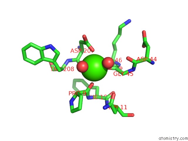
Mono view
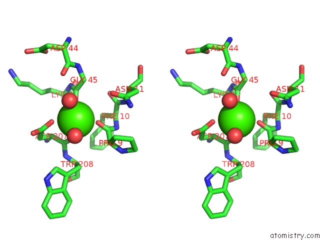
Stereo pair view
Mono view
Stereo pair view
A full contact list of Calcium with other atoms in the Ca binding
site number 1 of Crystal and Molecular Structure at 1.6 Angstroms Resolution of the Hybrid Bacillus Endo-1,3-1,4-Beta-D-Glucan 4- Glucanohydrolase H(A16-M) within 5.0Å range:
|
Reference:
M.Hahn,
T.Keitel,
U.Heinemann.
Crystal and Molecular Structure at 0.16-Nm Resolution of the Hybrid Bacillus Endo-1,3-1,4-Beta-D-Glucan 4-Glucanohydrolase H(A16-M). Eur.J.Biochem. V. 232 849 1995.
ISSN: ISSN 0014-2956
PubMed: 7588726
DOI: 10.1111/J.1432-1033.1995.TB20883.X
Page generated: Fri Jul 12 08:53:13 2024
ISSN: ISSN 0014-2956
PubMed: 7588726
DOI: 10.1111/J.1432-1033.1995.TB20883.X
Last articles
Zn in 9JYWZn in 9IR4
Zn in 9IR3
Zn in 9GMX
Zn in 9GMW
Zn in 9JEJ
Zn in 9ERF
Zn in 9ERE
Zn in 9EGV
Zn in 9EGW