Calcium »
PDB 2bir-2bw1 »
2bir »
Calcium in PDB 2bir: Additivity of Substrate Binding in Ribonuclease T1 (Y42A Mutant)
Protein crystallography data
The structure of Additivity of Substrate Binding in Ribonuclease T1 (Y42A Mutant), PDB code: 2bir
was solved by
J.Doumen,
J.Steyaert,
S.Loverix,
with X-Ray Crystallography technique. A brief refinement statistics is given in the table below:
| Resolution Low / High (Å) | 10.00 / 2.30 |
| Space group | P 21 21 21 |
| Cell size a, b, c (Å), α, β, γ (°) | 49.610, 48.490, 40.610, 90.00, 90.00, 90.00 |
| R / Rfree (%) | 21.6 / 25.7 |
Calcium Binding Sites:
The binding sites of Calcium atom in the Additivity of Substrate Binding in Ribonuclease T1 (Y42A Mutant)
(pdb code 2bir). This binding sites where shown within
5.0 Angstroms radius around Calcium atom.
In total only one binding site of Calcium was determined in the Additivity of Substrate Binding in Ribonuclease T1 (Y42A Mutant), PDB code: 2bir:
In total only one binding site of Calcium was determined in the Additivity of Substrate Binding in Ribonuclease T1 (Y42A Mutant), PDB code: 2bir:
Calcium binding site 1 out of 1 in 2bir
Go back to
Calcium binding site 1 out
of 1 in the Additivity of Substrate Binding in Ribonuclease T1 (Y42A Mutant)
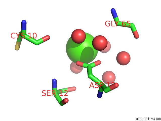
Mono view
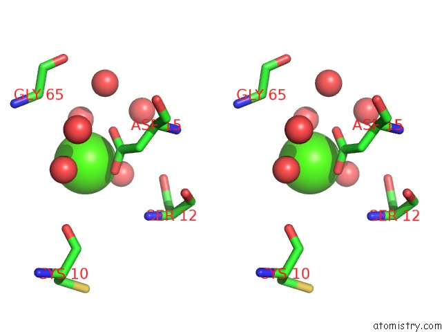
Stereo pair view
Mono view
Stereo pair view
A full contact list of Calcium with other atoms in the Ca binding
site number 1 of Additivity of Substrate Binding in Ribonuclease T1 (Y42A Mutant) within 5.0Å range:
|
Reference:
S.Loverix,
J.Doumen,
J.Steyaert.
Additivity of Protein-Guanine Interactions in Ribonuclease T1. J.Biol.Chem. V. 272 9635 1997.
ISSN: ISSN 0021-9258
PubMed: 9092491
DOI: 10.1074/JBC.272.15.9635
Page generated: Fri Jul 12 09:04:51 2024
ISSN: ISSN 0021-9258
PubMed: 9092491
DOI: 10.1074/JBC.272.15.9635
Last articles
Zn in 9J0NZn in 9J0O
Zn in 9J0P
Zn in 9FJX
Zn in 9EKB
Zn in 9C0F
Zn in 9CAH
Zn in 9CH0
Zn in 9CH3
Zn in 9CH1