Calcium »
PDB 2cn6-2dbx »
2d0v »
Calcium in PDB 2d0v: Crystal Structure of Methanol Dehydrogenase From Hyphomicrobium Denitrificans
Enzymatic activity of Crystal Structure of Methanol Dehydrogenase From Hyphomicrobium Denitrificans
All present enzymatic activity of Crystal Structure of Methanol Dehydrogenase From Hyphomicrobium Denitrificans:
1.1.99.8;
1.1.99.8;
Protein crystallography data
The structure of Crystal Structure of Methanol Dehydrogenase From Hyphomicrobium Denitrificans, PDB code: 2d0v
was solved by
M.Nojiri,
D.Hira,
K.Yamaguchi,
S.Suzuki,
with X-Ray Crystallography technique. A brief refinement statistics is given in the table below:
| Resolution Low / High (Å) | 44.60 / 2.49 |
| Space group | C 1 2 1 |
| Cell size a, b, c (Å), α, β, γ (°) | 291.320, 63.999, 109.941, 90.00, 105.74, 90.00 |
| R / Rfree (%) | 14.9 / 24.7 |
Calcium Binding Sites:
The binding sites of Calcium atom in the Crystal Structure of Methanol Dehydrogenase From Hyphomicrobium Denitrificans
(pdb code 2d0v). This binding sites where shown within
5.0 Angstroms radius around Calcium atom.
In total 3 binding sites of Calcium where determined in the Crystal Structure of Methanol Dehydrogenase From Hyphomicrobium Denitrificans, PDB code: 2d0v:
Jump to Calcium binding site number: 1; 2; 3;
In total 3 binding sites of Calcium where determined in the Crystal Structure of Methanol Dehydrogenase From Hyphomicrobium Denitrificans, PDB code: 2d0v:
Jump to Calcium binding site number: 1; 2; 3;
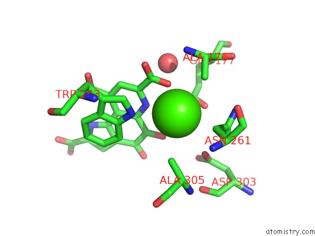
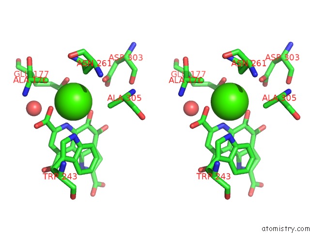
Calcium binding site 1 out of 3 in 2d0v
Go back to
Calcium binding site 1 out
of 3 in the Crystal Structure of Methanol Dehydrogenase From Hyphomicrobium Denitrificans
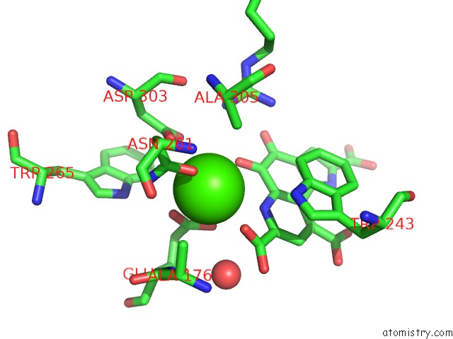
Mono view
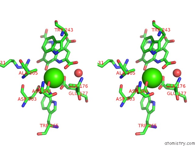
Stereo pair view
Mono view
Stereo pair view
A full contact list of Calcium with other atoms in the Ca binding
site number 1 of Crystal Structure of Methanol Dehydrogenase From Hyphomicrobium Denitrificans within 5.0Å range:
|
Calcium binding site 2 out of 3 in 2d0v
Go back to
Calcium binding site 2 out
of 3 in the Crystal Structure of Methanol Dehydrogenase From Hyphomicrobium Denitrificans
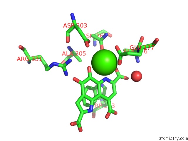
Mono view
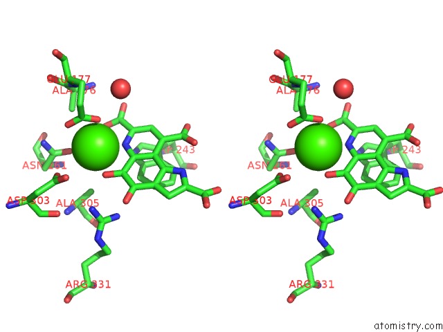
Stereo pair view
Mono view
Stereo pair view
A full contact list of Calcium with other atoms in the Ca binding
site number 2 of Crystal Structure of Methanol Dehydrogenase From Hyphomicrobium Denitrificans within 5.0Å range:
|
Calcium binding site 3 out of 3 in 2d0v
Go back to
Calcium binding site 3 out
of 3 in the Crystal Structure of Methanol Dehydrogenase From Hyphomicrobium Denitrificans
Mono view
Stereo pair view
Mono view
Stereo pair view
A full contact list of Calcium with other atoms in the Ca binding
site number 3 of Crystal Structure of Methanol Dehydrogenase From Hyphomicrobium Denitrificans within 5.0Å range:
|
Reference:
M.Nojiri,
D.Hira,
K.Yamaguchi,
T.Okajima,
K.Tanizawa,
S.Suzuki.
Crystal Structures of Cytochrome C(L) and Methanol Dehydrogenase From Hyphomicrobium Denitrificans: Structural and Mechanistic Insights Into Interactions Between the Two Proteins Biochemistry V. 45 3481 2006.
ISSN: ISSN 0006-2960
PubMed: 16533029
DOI: 10.1021/BI051877J
Page generated: Fri Jul 12 09:38:44 2024
ISSN: ISSN 0006-2960
PubMed: 16533029
DOI: 10.1021/BI051877J
Last articles
Zn in 9MJ5Zn in 9HNW
Zn in 9G0L
Zn in 9FNE
Zn in 9DZN
Zn in 9E0I
Zn in 9D32
Zn in 9DAK
Zn in 8ZXC
Zn in 8ZUF