Calcium »
PDB 2cn6-2dbx »
2d4c »
Calcium in PDB 2d4c: Crystal Structure of the Endophilin Bar Domain Mutant
Protein crystallography data
The structure of Crystal Structure of the Endophilin Bar Domain Mutant, PDB code: 2d4c
was solved by
M.Masuda,
S.Takeda,
with X-Ray Crystallography technique. A brief refinement statistics is given in the table below:
| Resolution Low / High (Å) | 50.00 / 2.40 |
| Space group | P 1 21 1 |
| Cell size a, b, c (Å), α, β, γ (°) | 44.344, 212.217, 54.317, 90.00, 96.97, 90.00 |
| R / Rfree (%) | 21.5 / 27 |
Calcium Binding Sites:
The binding sites of Calcium atom in the Crystal Structure of the Endophilin Bar Domain Mutant
(pdb code 2d4c). This binding sites where shown within
5.0 Angstroms radius around Calcium atom.
In total only one binding site of Calcium was determined in the Crystal Structure of the Endophilin Bar Domain Mutant, PDB code: 2d4c:
In total only one binding site of Calcium was determined in the Crystal Structure of the Endophilin Bar Domain Mutant, PDB code: 2d4c:
Calcium binding site 1 out of 1 in 2d4c
Go back to
Calcium binding site 1 out
of 1 in the Crystal Structure of the Endophilin Bar Domain Mutant

Mono view
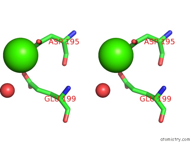
Stereo pair view
Mono view
Stereo pair view
A full contact list of Calcium with other atoms in the Ca binding
site number 1 of Crystal Structure of the Endophilin Bar Domain Mutant within 5.0Å range:
|
Reference:
M.Masuda,
S.Takeda,
M.Sone,
T.Ohki,
H.Mori,
Y.Kamioka,
N.Mochizuki.
Endophilin Bar Domain Drives Membrane Curvature By Two Newly Identified Structure-Based Mechanisms Embo J. V. 25 2889 2006.
ISSN: ISSN 0261-4189
PubMed: 16763557
DOI: 10.1038/SJ.EMBOJ.7601176
Page generated: Fri Jul 12 09:43:10 2024
ISSN: ISSN 0261-4189
PubMed: 16763557
DOI: 10.1038/SJ.EMBOJ.7601176
Last articles
Zn in 9J0NZn in 9J0O
Zn in 9J0P
Zn in 9FJX
Zn in 9EKB
Zn in 9C0F
Zn in 9CAH
Zn in 9CH0
Zn in 9CH3
Zn in 9CH1