Calcium »
PDB 2cn6-2dbx »
2d7f »
Calcium in PDB 2d7f: Crystal Structure of A Lectin From Canavalia Gladiata Seeds Complexed with Alpha-Methyl-Mannoside and Alpha- Aminobutyric Acid
Protein crystallography data
The structure of Crystal Structure of A Lectin From Canavalia Gladiata Seeds Complexed with Alpha-Methyl-Mannoside and Alpha- Aminobutyric Acid, PDB code: 2d7f
was solved by
P.Delatorre,
B.A.M.Rocha,
E.P.Souza,
B.T.Freitas,
F.B.B.M.Moreno,
A.H.Sampaio,
W.F.Azevedo Jr.,
B.S.Cavada,
with X-Ray Crystallography technique. A brief refinement statistics is given in the table below:
| Resolution Low / High (Å) | 9.99 / 2.31 |
| Space group | C 2 2 21 |
| Cell size a, b, c (Å), α, β, γ (°) | 100.915, 115.754, 241.626, 90.00, 90.00, 90.00 |
| R / Rfree (%) | 18 / 22.7 |
Other elements in 2d7f:
The structure of Crystal Structure of A Lectin From Canavalia Gladiata Seeds Complexed with Alpha-Methyl-Mannoside and Alpha- Aminobutyric Acid also contains other interesting chemical elements:
| Manganese | (Mn) | 4 atoms |
Calcium Binding Sites:
The binding sites of Calcium atom in the Crystal Structure of A Lectin From Canavalia Gladiata Seeds Complexed with Alpha-Methyl-Mannoside and Alpha- Aminobutyric Acid
(pdb code 2d7f). This binding sites where shown within
5.0 Angstroms radius around Calcium atom.
In total 4 binding sites of Calcium where determined in the Crystal Structure of A Lectin From Canavalia Gladiata Seeds Complexed with Alpha-Methyl-Mannoside and Alpha- Aminobutyric Acid, PDB code: 2d7f:
Jump to Calcium binding site number: 1; 2; 3; 4;
In total 4 binding sites of Calcium where determined in the Crystal Structure of A Lectin From Canavalia Gladiata Seeds Complexed with Alpha-Methyl-Mannoside and Alpha- Aminobutyric Acid, PDB code: 2d7f:
Jump to Calcium binding site number: 1; 2; 3; 4;
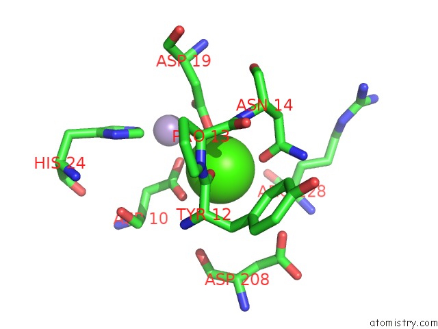
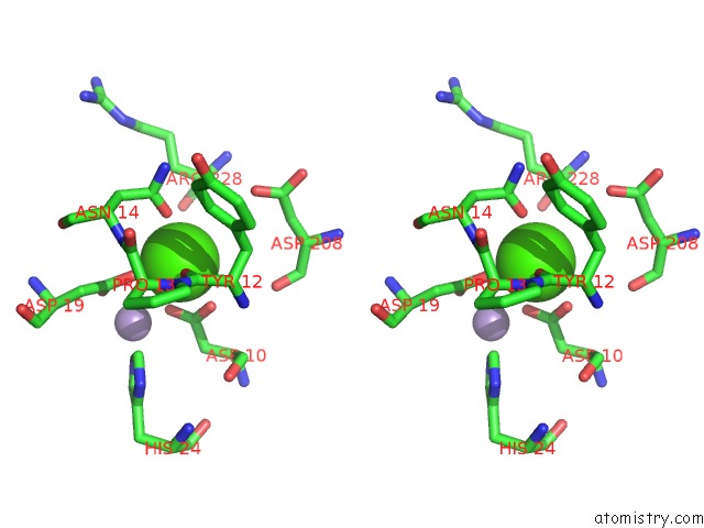
Calcium binding site 1 out of 4 in 2d7f
Go back to
Calcium binding site 1 out
of 4 in the Crystal Structure of A Lectin From Canavalia Gladiata Seeds Complexed with Alpha-Methyl-Mannoside and Alpha- Aminobutyric Acid
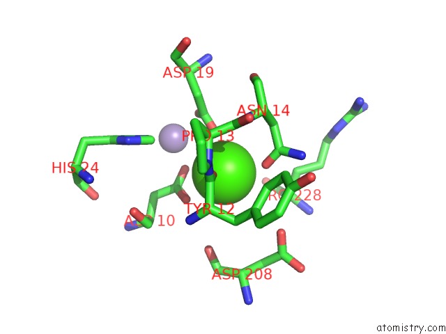
Mono view
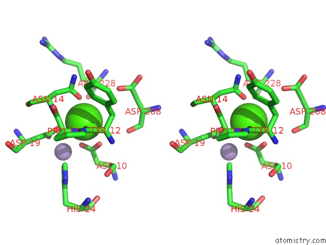
Stereo pair view
Mono view
Stereo pair view
A full contact list of Calcium with other atoms in the Ca binding
site number 1 of Crystal Structure of A Lectin From Canavalia Gladiata Seeds Complexed with Alpha-Methyl-Mannoside and Alpha- Aminobutyric Acid within 5.0Å range:
|
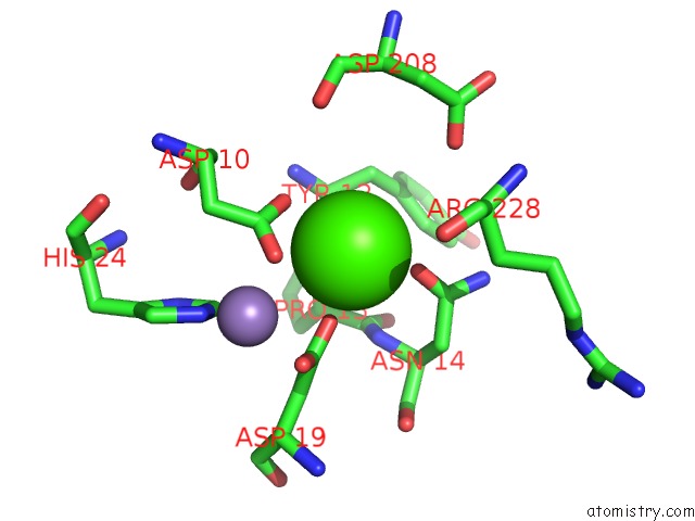
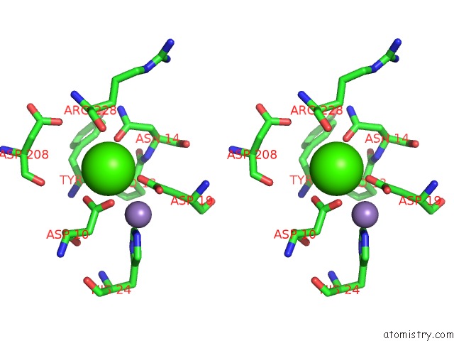
Calcium binding site 2 out of 4 in 2d7f
Go back to
Calcium binding site 2 out
of 4 in the Crystal Structure of A Lectin From Canavalia Gladiata Seeds Complexed with Alpha-Methyl-Mannoside and Alpha- Aminobutyric Acid
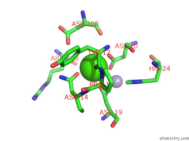
Mono view
Stereo pair view
Mono view
Stereo pair view
A full contact list of Calcium with other atoms in the Ca binding
site number 2 of Crystal Structure of A Lectin From Canavalia Gladiata Seeds Complexed with Alpha-Methyl-Mannoside and Alpha- Aminobutyric Acid within 5.0Å range:
|
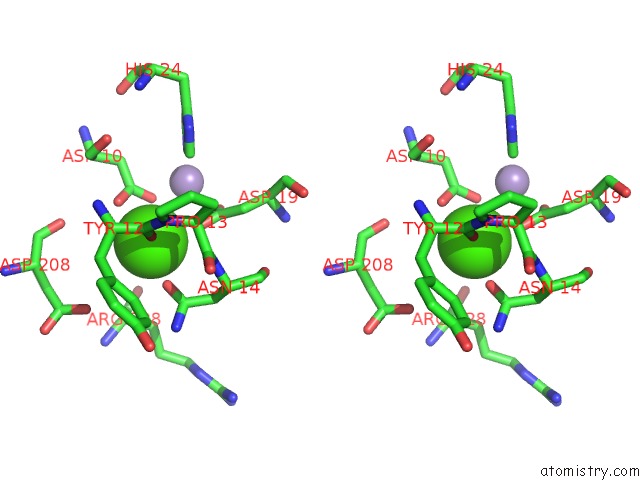
Calcium binding site 3 out of 4 in 2d7f
Go back to
Calcium binding site 3 out
of 4 in the Crystal Structure of A Lectin From Canavalia Gladiata Seeds Complexed with Alpha-Methyl-Mannoside and Alpha- Aminobutyric Acid
Mono view
Stereo pair view
Mono view
Stereo pair view
A full contact list of Calcium with other atoms in the Ca binding
site number 3 of Crystal Structure of A Lectin From Canavalia Gladiata Seeds Complexed with Alpha-Methyl-Mannoside and Alpha- Aminobutyric Acid within 5.0Å range:
|
Calcium binding site 4 out of 4 in 2d7f
Go back to
Calcium binding site 4 out
of 4 in the Crystal Structure of A Lectin From Canavalia Gladiata Seeds Complexed with Alpha-Methyl-Mannoside and Alpha- Aminobutyric Acid
Mono view
Stereo pair view
Mono view
Stereo pair view
A full contact list of Calcium with other atoms in the Ca binding
site number 4 of Crystal Structure of A Lectin From Canavalia Gladiata Seeds Complexed with Alpha-Methyl-Mannoside and Alpha- Aminobutyric Acid within 5.0Å range:
|
Reference:
P.Delatorre,
B.A.M.Rocha,
E.P.Souza,
T.M.Oliveira,
G.A.Bezerra,
F.B.M.B.Moreno,
B.T.Freitas,
T.Santi-Gadelha,
A.H.Sampaio,
W.F.Azevedo Jr.,
B.S.Cavada.
Structure of A Lectin From Canavalia Gladiata Seeds: New Structural Insights For Old Molecules Bmc Struct.Biol. V. 7 52 2007.
ISSN: ESSN 1472-6807
PubMed: 17683532
DOI: 10.1186/1472-6807-7-52
Page generated: Fri Jul 12 09:43:39 2024
ISSN: ESSN 1472-6807
PubMed: 17683532
DOI: 10.1186/1472-6807-7-52
Last articles
Zn in 9J0NZn in 9J0O
Zn in 9J0P
Zn in 9FJX
Zn in 9EKB
Zn in 9C0F
Zn in 9CAH
Zn in 9CH0
Zn in 9CH3
Zn in 9CH1