Calcium »
PDB 2dcj-2dw0 »
2ddu »
Calcium in PDB 2ddu: Crystal Structure of the Third Repeat Domain of Reelin
Protein crystallography data
The structure of Crystal Structure of the Third Repeat Domain of Reelin, PDB code: 2ddu
was solved by
T.Nogi,
N.Yasui,
J.Takagi,
with X-Ray Crystallography technique. A brief refinement statistics is given in the table below:
| Resolution Low / High (Å) | 50.00 / 2.05 |
| Space group | H 3 2 |
| Cell size a, b, c (Å), α, β, γ (°) | 129.932, 129.932, 122.485, 90.00, 90.00, 120.00 |
| R / Rfree (%) | 23.4 / 26.5 |
Other elements in 2ddu:
The structure of Crystal Structure of the Third Repeat Domain of Reelin also contains other interesting chemical elements:
| Magnesium | (Mg) | 1 atom |
| Chlorine | (Cl) | 1 atom |
Calcium Binding Sites:
The binding sites of Calcium atom in the Crystal Structure of the Third Repeat Domain of Reelin
(pdb code 2ddu). This binding sites where shown within
5.0 Angstroms radius around Calcium atom.
In total only one binding site of Calcium was determined in the Crystal Structure of the Third Repeat Domain of Reelin, PDB code: 2ddu:
In total only one binding site of Calcium was determined in the Crystal Structure of the Third Repeat Domain of Reelin, PDB code: 2ddu:
Calcium binding site 1 out of 1 in 2ddu
Go back to
Calcium binding site 1 out
of 1 in the Crystal Structure of the Third Repeat Domain of Reelin
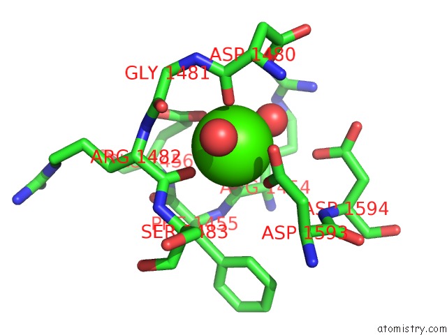
Mono view
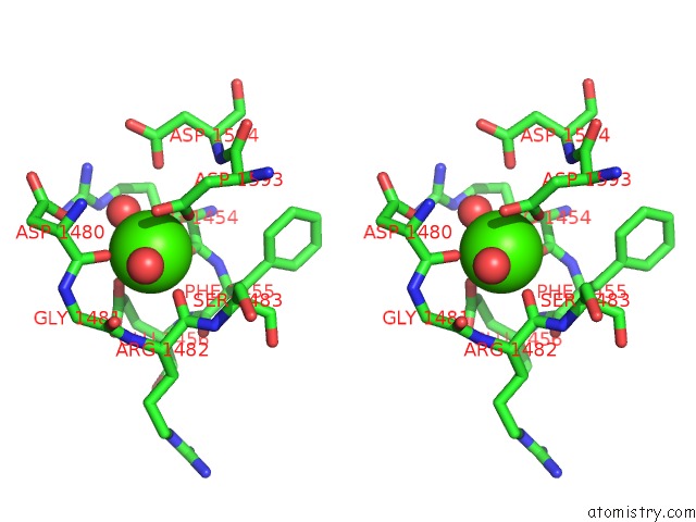
Stereo pair view
Mono view
Stereo pair view
A full contact list of Calcium with other atoms in the Ca binding
site number 1 of Crystal Structure of the Third Repeat Domain of Reelin within 5.0Å range:
|
Reference:
T.Nogi,
N.Yasui,
M.Hattori,
K.Iwasaki,
J.Takagi.
Structure of A Signaling-Competent Reelin Fragment Revealed By X-Ray Crystallography and Electron Tomography Embo J. V. 25 3675 2006.
ISSN: ISSN 0261-4189
PubMed: 16858396
DOI: 10.1038/SJ.EMBOJ.7601240
Page generated: Fri Jul 12 09:45:04 2024
ISSN: ISSN 0261-4189
PubMed: 16858396
DOI: 10.1038/SJ.EMBOJ.7601240
Last articles
Zn in 9J0NZn in 9J0O
Zn in 9J0P
Zn in 9FJX
Zn in 9EKB
Zn in 9C0F
Zn in 9CAH
Zn in 9CH0
Zn in 9CH3
Zn in 9CH1