Calcium »
PDB 2dcj-2dw0 »
2duo »
Calcium in PDB 2duo: Crystal Structure of VIP36 Exoplasmic/Lumenal Domain, CA2+- Bound Form
Protein crystallography data
The structure of Crystal Structure of VIP36 Exoplasmic/Lumenal Domain, CA2+- Bound Form, PDB code: 2duo
was solved by
T.Satoh,
N.P.Cowieson,
R.Kato,
S.Wakatsuki,
with X-Ray Crystallography technique. A brief refinement statistics is given in the table below:
| Resolution Low / High (Å) | 20.00 / 1.80 |
| Space group | C 1 2 1 |
| Cell size a, b, c (Å), α, β, γ (°) | 170.700, 45.400, 116.100, 90.00, 131.50, 90.00 |
| R / Rfree (%) | 20.5 / 24.1 |
Other elements in 2duo:
The structure of Crystal Structure of VIP36 Exoplasmic/Lumenal Domain, CA2+- Bound Form also contains other interesting chemical elements:
| Chlorine | (Cl) | 11 atoms |
Calcium Binding Sites:
The binding sites of Calcium atom in the Crystal Structure of VIP36 Exoplasmic/Lumenal Domain, CA2+- Bound Form
(pdb code 2duo). This binding sites where shown within
5.0 Angstroms radius around Calcium atom.
In total 2 binding sites of Calcium where determined in the Crystal Structure of VIP36 Exoplasmic/Lumenal Domain, CA2+- Bound Form, PDB code: 2duo:
Jump to Calcium binding site number: 1; 2;
In total 2 binding sites of Calcium where determined in the Crystal Structure of VIP36 Exoplasmic/Lumenal Domain, CA2+- Bound Form, PDB code: 2duo:
Jump to Calcium binding site number: 1; 2;
Calcium binding site 1 out of 2 in 2duo
Go back to
Calcium binding site 1 out
of 2 in the Crystal Structure of VIP36 Exoplasmic/Lumenal Domain, CA2+- Bound Form
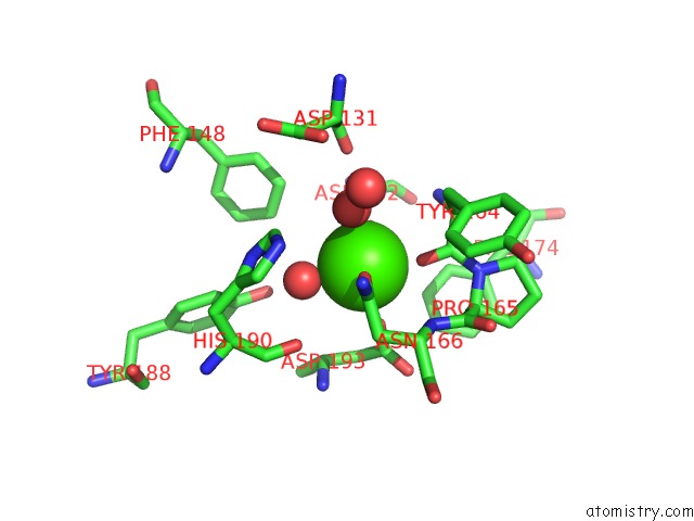
Mono view
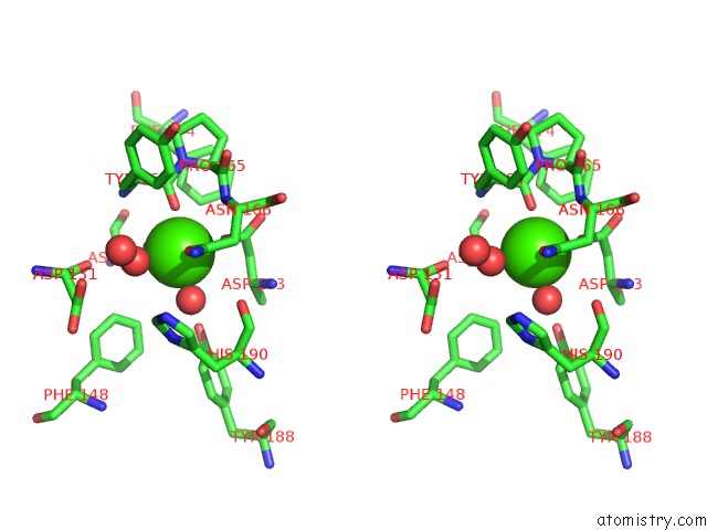
Stereo pair view
Mono view
Stereo pair view
A full contact list of Calcium with other atoms in the Ca binding
site number 1 of Crystal Structure of VIP36 Exoplasmic/Lumenal Domain, CA2+- Bound Form within 5.0Å range:
|
Calcium binding site 2 out of 2 in 2duo
Go back to
Calcium binding site 2 out
of 2 in the Crystal Structure of VIP36 Exoplasmic/Lumenal Domain, CA2+- Bound Form
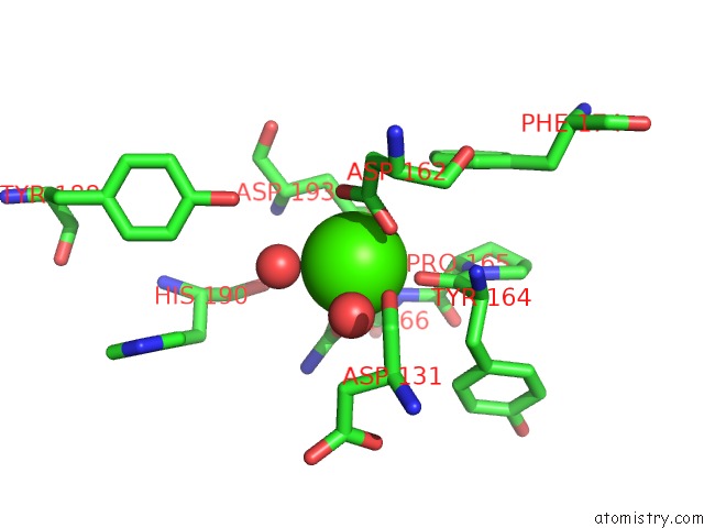
Mono view
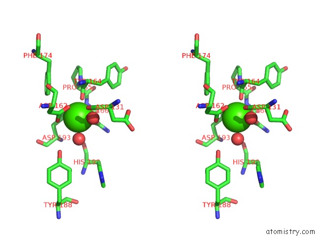
Stereo pair view
Mono view
Stereo pair view
A full contact list of Calcium with other atoms in the Ca binding
site number 2 of Crystal Structure of VIP36 Exoplasmic/Lumenal Domain, CA2+- Bound Form within 5.0Å range:
|
Reference:
T.Satoh,
N.P.Cowieson,
W.Hakamata,
H.Ideo,
K.Fukushima,
M.Kurihara,
R.Kato,
K.Yamashita,
S.Wakatsuki.
Structural Basis For Recognition of High Mannose Type Glycoproteins By Mammalian Transport Lectin VIP36 J.Biol.Chem. V. 282 28246 2007.
ISSN: ISSN 0021-9258
PubMed: 17652092
DOI: 10.1074/JBC.M703064200
Page generated: Fri Jul 12 09:54:15 2024
ISSN: ISSN 0021-9258
PubMed: 17652092
DOI: 10.1074/JBC.M703064200
Last articles
Zn in 9J0NZn in 9J0O
Zn in 9J0P
Zn in 9FJX
Zn in 9EKB
Zn in 9C0F
Zn in 9CAH
Zn in 9CH0
Zn in 9CH3
Zn in 9CH1