Calcium »
PDB 2dcj-2dw0 »
2dvf »
Calcium in PDB 2dvf: Crystals of Peanut Lectin Grown in the Presence of Gal- Alpha-1,3-Gal-Beta-1,4-Gal
Protein crystallography data
The structure of Crystals of Peanut Lectin Grown in the Presence of Gal- Alpha-1,3-Gal-Beta-1,4-Gal, PDB code: 2dvf
was solved by
S.K.Natchiar,
O.Srinivas,
N.Mitra,
A.Surolia,
N.Jayaraman,
M.Vijayan,
with X-Ray Crystallography technique. A brief refinement statistics is given in the table below:
| Resolution Low / High (Å) | 19.94 / 2.74 |
| Space group | P 21 21 2 |
| Cell size a, b, c (Å), α, β, γ (°) | 125.810, 124.060, 75.400, 90.00, 90.00, 90.00 |
| R / Rfree (%) | 20 / 26.3 |
Other elements in 2dvf:
The structure of Crystals of Peanut Lectin Grown in the Presence of Gal- Alpha-1,3-Gal-Beta-1,4-Gal also contains other interesting chemical elements:
| Manganese | (Mn) | 4 atoms |
Calcium Binding Sites:
The binding sites of Calcium atom in the Crystals of Peanut Lectin Grown in the Presence of Gal- Alpha-1,3-Gal-Beta-1,4-Gal
(pdb code 2dvf). This binding sites where shown within
5.0 Angstroms radius around Calcium atom.
In total 4 binding sites of Calcium where determined in the Crystals of Peanut Lectin Grown in the Presence of Gal- Alpha-1,3-Gal-Beta-1,4-Gal, PDB code: 2dvf:
Jump to Calcium binding site number: 1; 2; 3; 4;
In total 4 binding sites of Calcium where determined in the Crystals of Peanut Lectin Grown in the Presence of Gal- Alpha-1,3-Gal-Beta-1,4-Gal, PDB code: 2dvf:
Jump to Calcium binding site number: 1; 2; 3; 4;
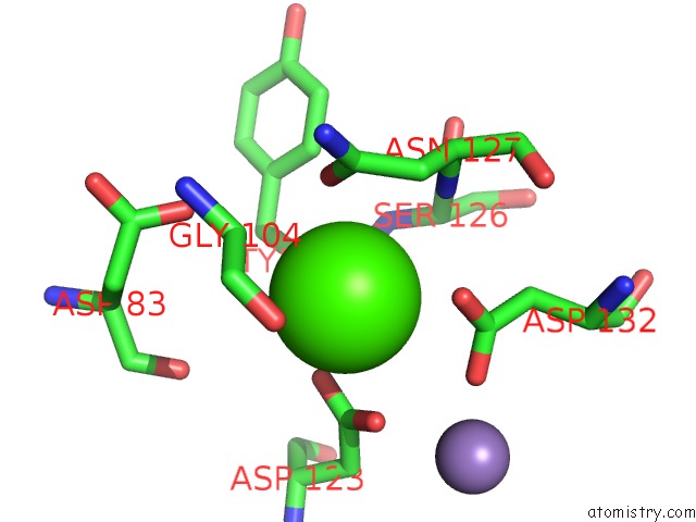
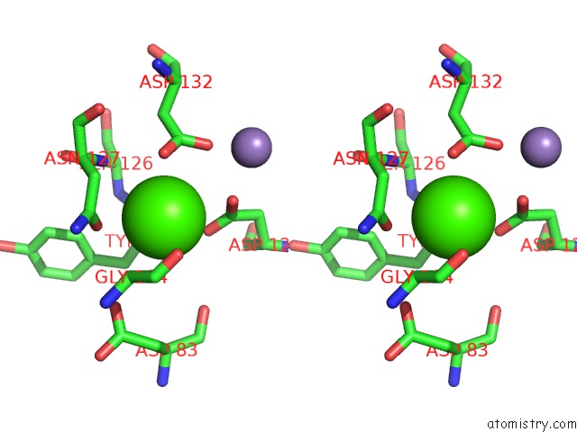
Calcium binding site 1 out of 4 in 2dvf
Go back to
Calcium binding site 1 out
of 4 in the Crystals of Peanut Lectin Grown in the Presence of Gal- Alpha-1,3-Gal-Beta-1,4-Gal
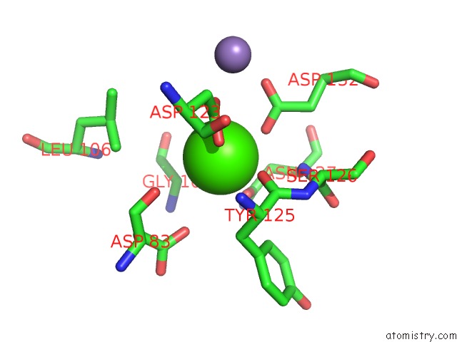
Mono view
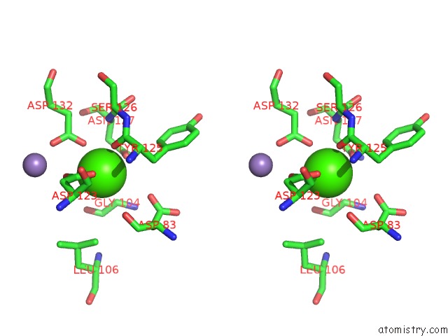
Stereo pair view
Mono view
Stereo pair view
A full contact list of Calcium with other atoms in the Ca binding
site number 1 of Crystals of Peanut Lectin Grown in the Presence of Gal- Alpha-1,3-Gal-Beta-1,4-Gal within 5.0Å range:
|
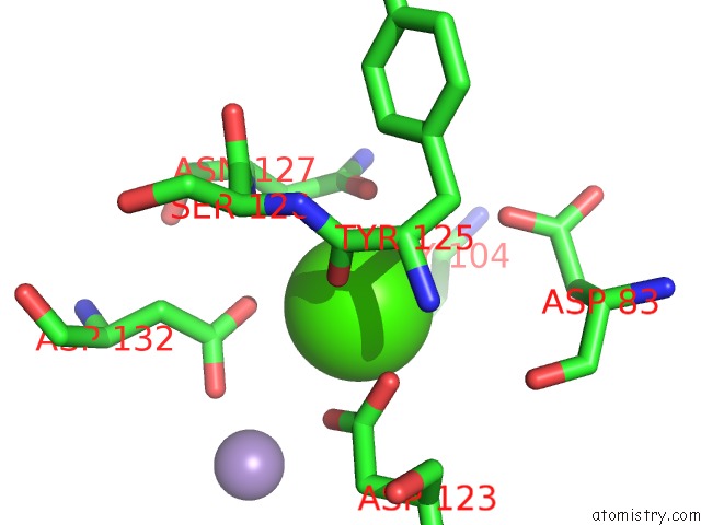
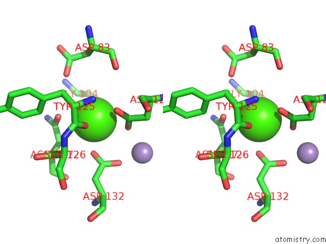
Calcium binding site 2 out of 4 in 2dvf
Go back to
Calcium binding site 2 out
of 4 in the Crystals of Peanut Lectin Grown in the Presence of Gal- Alpha-1,3-Gal-Beta-1,4-Gal
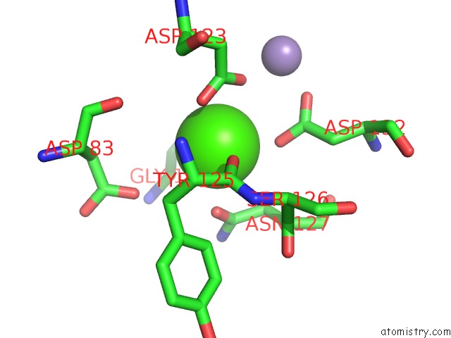
Mono view
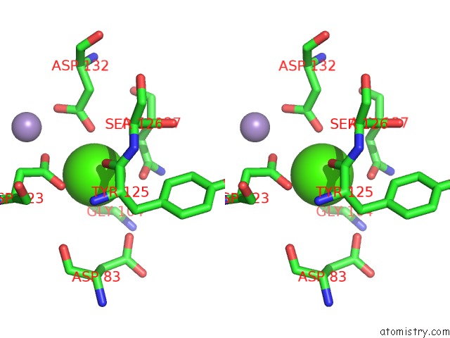
Stereo pair view
Mono view
Stereo pair view
A full contact list of Calcium with other atoms in the Ca binding
site number 2 of Crystals of Peanut Lectin Grown in the Presence of Gal- Alpha-1,3-Gal-Beta-1,4-Gal within 5.0Å range:
|
Calcium binding site 3 out of 4 in 2dvf
Go back to
Calcium binding site 3 out
of 4 in the Crystals of Peanut Lectin Grown in the Presence of Gal- Alpha-1,3-Gal-Beta-1,4-Gal
Mono view
Stereo pair view
Mono view
Stereo pair view
A full contact list of Calcium with other atoms in the Ca binding
site number 3 of Crystals of Peanut Lectin Grown in the Presence of Gal- Alpha-1,3-Gal-Beta-1,4-Gal within 5.0Å range:
|
Calcium binding site 4 out of 4 in 2dvf
Go back to
Calcium binding site 4 out
of 4 in the Crystals of Peanut Lectin Grown in the Presence of Gal- Alpha-1,3-Gal-Beta-1,4-Gal
Mono view
Stereo pair view
Mono view
Stereo pair view
A full contact list of Calcium with other atoms in the Ca binding
site number 4 of Crystals of Peanut Lectin Grown in the Presence of Gal- Alpha-1,3-Gal-Beta-1,4-Gal within 5.0Å range:
|
Reference:
S.K.Natchiar,
O.Srinivas,
N.Mitra,
A.Surolia,
N.Jayaraman,
M.Vijayan.
Structural Studies on Peanut Lectin Complexed with Disaccharides Involving Different Linkages: Further Insights Into the Structure and Interactions of the Lectin Acta Crystallogr.,Sect.D V. 62 1413 2006.
ISSN: ISSN 0907-4449
PubMed: 17057347
DOI: 10.1107/S0907444906035712
Page generated: Fri Jul 12 09:56:05 2024
ISSN: ISSN 0907-4449
PubMed: 17057347
DOI: 10.1107/S0907444906035712
Last articles
Zn in 9MJ5Zn in 9HNW
Zn in 9G0L
Zn in 9FNE
Zn in 9DZN
Zn in 9E0I
Zn in 9D32
Zn in 9DAK
Zn in 8ZXC
Zn in 8ZUF