Calcium »
PDB 2dw1-2eek »
2e3t »
Calcium in PDB 2e3t: Crystal Structure of Rat Xanthine Oxidoreductase Mutant (W335A and F336L)
Enzymatic activity of Crystal Structure of Rat Xanthine Oxidoreductase Mutant (W335A and F336L)
All present enzymatic activity of Crystal Structure of Rat Xanthine Oxidoreductase Mutant (W335A and F336L):
1.17.1.4; 1.17.3.2;
1.17.1.4; 1.17.3.2;
Protein crystallography data
The structure of Crystal Structure of Rat Xanthine Oxidoreductase Mutant (W335A and F336L), PDB code: 2e3t
was solved by
R.Asai,
T.Nishino,
T.Matsumura,
K.Okamoto,
E.F.Pai,
T.Nishino,
with X-Ray Crystallography technique. A brief refinement statistics is given in the table below:
| Resolution Low / High (Å) | 25.00 / 2.28 |
| Space group | P 21 21 21 |
| Cell size a, b, c (Å), α, β, γ (°) | 101.066, 139.513, 222.304, 90.00, 90.00, 90.00 |
| R / Rfree (%) | 18.5 / 22.9 |
Other elements in 2e3t:
The structure of Crystal Structure of Rat Xanthine Oxidoreductase Mutant (W335A and F336L) also contains other interesting chemical elements:
| Iron | (Fe) | 8 atoms |
Calcium Binding Sites:
The binding sites of Calcium atom in the Crystal Structure of Rat Xanthine Oxidoreductase Mutant (W335A and F336L)
(pdb code 2e3t). This binding sites where shown within
5.0 Angstroms radius around Calcium atom.
In total 4 binding sites of Calcium where determined in the Crystal Structure of Rat Xanthine Oxidoreductase Mutant (W335A and F336L), PDB code: 2e3t:
Jump to Calcium binding site number: 1; 2; 3; 4;
In total 4 binding sites of Calcium where determined in the Crystal Structure of Rat Xanthine Oxidoreductase Mutant (W335A and F336L), PDB code: 2e3t:
Jump to Calcium binding site number: 1; 2; 3; 4;
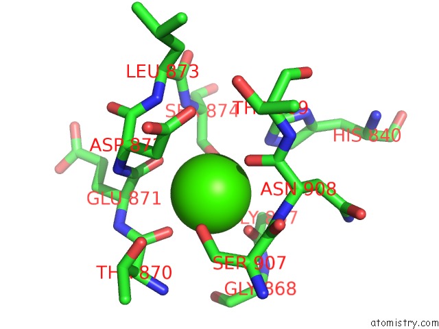
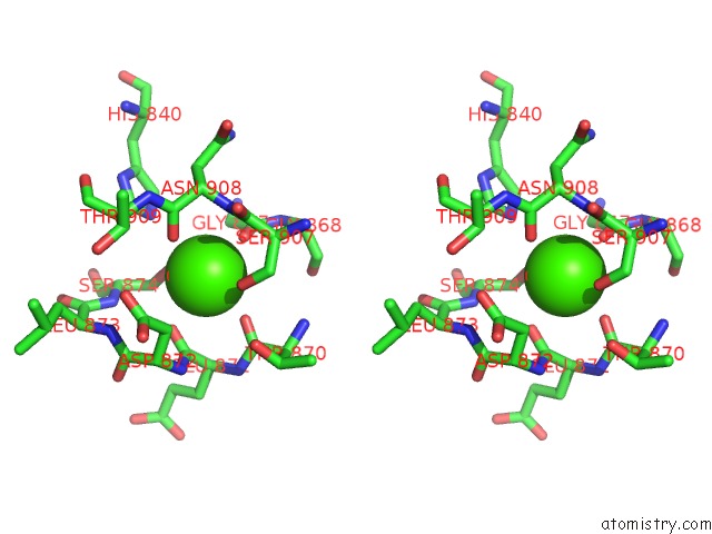
Calcium binding site 1 out of 4 in 2e3t
Go back to
Calcium binding site 1 out
of 4 in the Crystal Structure of Rat Xanthine Oxidoreductase Mutant (W335A and F336L)
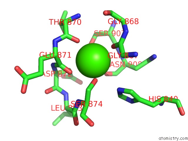
Mono view
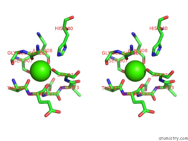
Stereo pair view
Mono view
Stereo pair view
A full contact list of Calcium with other atoms in the Ca binding
site number 1 of Crystal Structure of Rat Xanthine Oxidoreductase Mutant (W335A and F336L) within 5.0Å range:
|
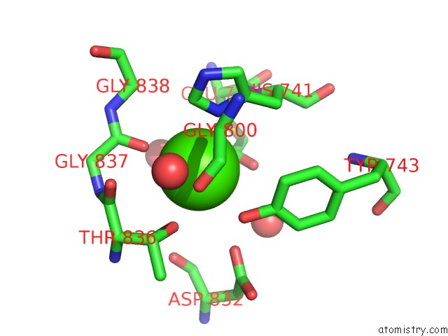
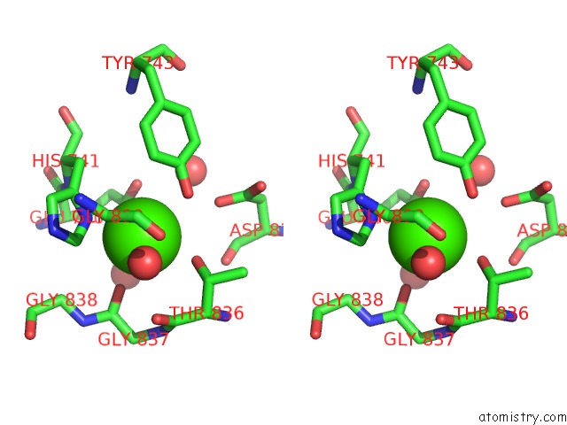
Calcium binding site 2 out of 4 in 2e3t
Go back to
Calcium binding site 2 out
of 4 in the Crystal Structure of Rat Xanthine Oxidoreductase Mutant (W335A and F336L)
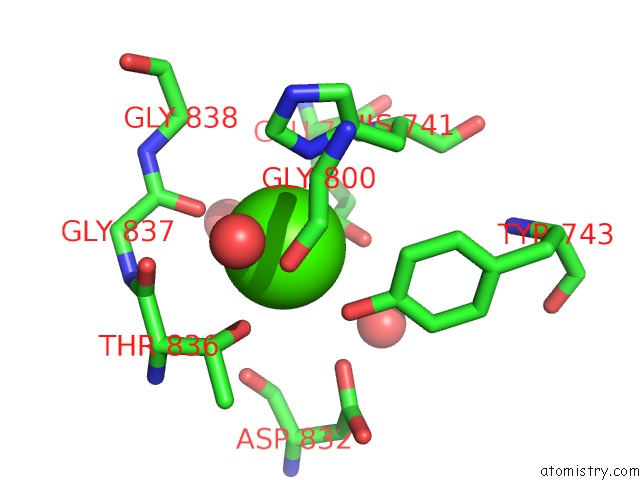
Mono view
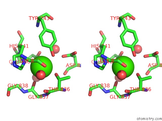
Stereo pair view
Mono view
Stereo pair view
A full contact list of Calcium with other atoms in the Ca binding
site number 2 of Crystal Structure of Rat Xanthine Oxidoreductase Mutant (W335A and F336L) within 5.0Å range:
|
Calcium binding site 3 out of 4 in 2e3t
Go back to
Calcium binding site 3 out
of 4 in the Crystal Structure of Rat Xanthine Oxidoreductase Mutant (W335A and F336L)
Mono view
Stereo pair view
Mono view
Stereo pair view
A full contact list of Calcium with other atoms in the Ca binding
site number 3 of Crystal Structure of Rat Xanthine Oxidoreductase Mutant (W335A and F336L) within 5.0Å range:
|
Calcium binding site 4 out of 4 in 2e3t
Go back to
Calcium binding site 4 out
of 4 in the Crystal Structure of Rat Xanthine Oxidoreductase Mutant (W335A and F336L)
Mono view
Stereo pair view
Mono view
Stereo pair view
A full contact list of Calcium with other atoms in the Ca binding
site number 4 of Crystal Structure of Rat Xanthine Oxidoreductase Mutant (W335A and F336L) within 5.0Å range:
|
Reference:
R.Asai,
T.Nishino,
T.Matsumura,
K.Okamoto,
K.Igarashi,
E.F.Pai,
T.Nishino.
Two Mutations Convert Mammalian Xanthine Oxidoreductase to Highly Superoxide-Productive Xanthine Oxidase J.Biochem.(Tokyo) V. 141 525 2007.
ISSN: ISSN 0021-924X
PubMed: 17301076
DOI: 10.1093/JB/MVM054
Page generated: Tue Jul 8 05:14:56 2025
ISSN: ISSN 0021-924X
PubMed: 17301076
DOI: 10.1093/JB/MVM054
Last articles
F in 4EPXF in 4ENC
F in 4ENB
F in 4EMV
F in 4ENA
F in 4EN5
F in 4EKC
F in 4EKD
F in 4EHG
F in 4EHE