Calcium »
PDB 2dw1-2eek »
2e85 »
Calcium in PDB 2e85: Crystal Structure of the Hydrogenase 3 Maturation Protease
Protein crystallography data
The structure of Crystal Structure of the Hydrogenase 3 Maturation Protease, PDB code: 2e85
was solved by
T.Tanaka,
T.S.Kumarevel,
A.Shinkai,
S.Yokoyama,
Riken Structuralgenomics/Proteomics Initiative (Rsgi),
with X-Ray Crystallography technique. A brief refinement statistics is given in the table below:
| Resolution Low / High (Å) | 20.00 / 1.70 |
| Space group | P 21 21 2 |
| Cell size a, b, c (Å), α, β, γ (°) | 87.425, 54.984, 67.964, 90.00, 90.00, 90.00 |
| R / Rfree (%) | 24.8 / 25.8 |
Calcium Binding Sites:
The binding sites of Calcium atom in the Crystal Structure of the Hydrogenase 3 Maturation Protease
(pdb code 2e85). This binding sites where shown within
5.0 Angstroms radius around Calcium atom.
In total 6 binding sites of Calcium where determined in the Crystal Structure of the Hydrogenase 3 Maturation Protease, PDB code: 2e85:
Jump to Calcium binding site number: 1; 2; 3; 4; 5; 6;
In total 6 binding sites of Calcium where determined in the Crystal Structure of the Hydrogenase 3 Maturation Protease, PDB code: 2e85:
Jump to Calcium binding site number: 1; 2; 3; 4; 5; 6;
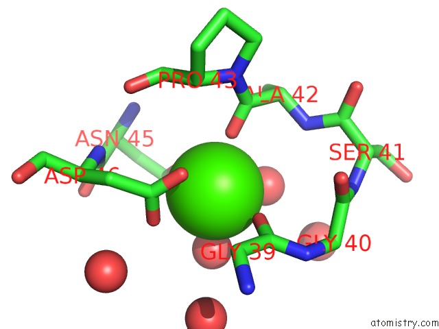
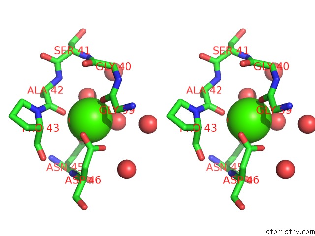
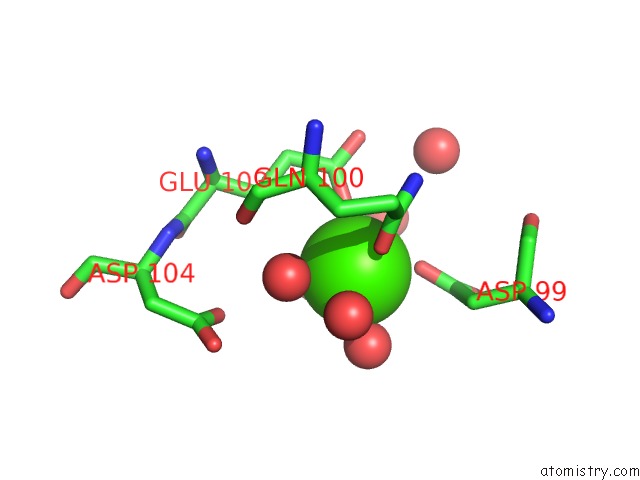
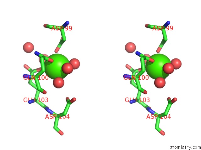
Calcium binding site 1 out of 6 in 2e85
Go back to
Calcium binding site 1 out
of 6 in the Crystal Structure of the Hydrogenase 3 Maturation Protease
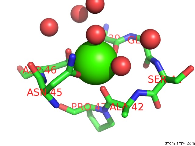
Mono view
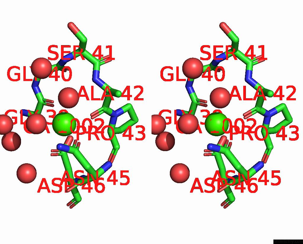
Stereo pair view
Mono view
Stereo pair view
A full contact list of Calcium with other atoms in the Ca binding
site number 1 of Crystal Structure of the Hydrogenase 3 Maturation Protease within 5.0Å range:
|
Calcium binding site 2 out of 6 in 2e85
Go back to
Calcium binding site 2 out
of 6 in the Crystal Structure of the Hydrogenase 3 Maturation Protease
Mono view
Stereo pair view
Mono view
Stereo pair view
A full contact list of Calcium with other atoms in the Ca binding
site number 2 of Crystal Structure of the Hydrogenase 3 Maturation Protease within 5.0Å range:
|
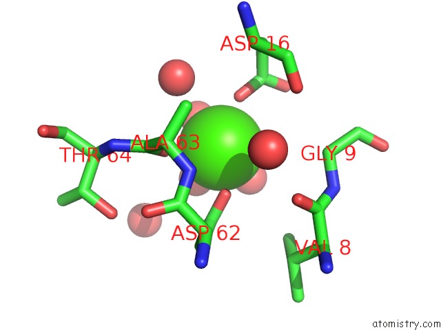
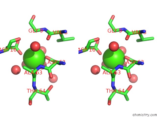
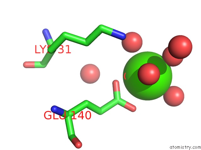
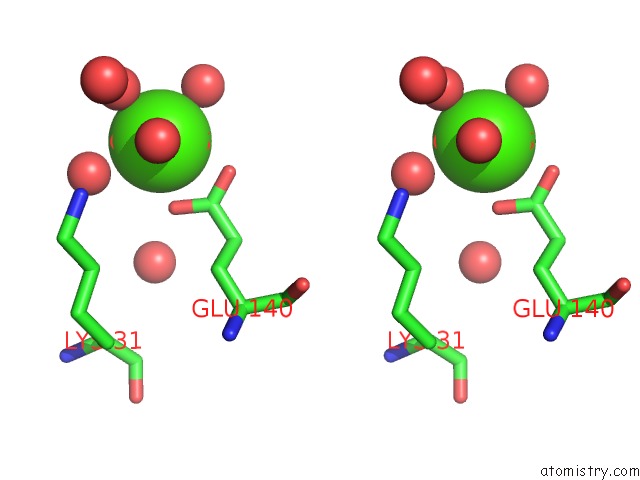
Calcium binding site 3 out of 6 in 2e85
Go back to
Calcium binding site 3 out
of 6 in the Crystal Structure of the Hydrogenase 3 Maturation Protease
Mono view
Stereo pair view
Mono view
Stereo pair view
A full contact list of Calcium with other atoms in the Ca binding
site number 3 of Crystal Structure of the Hydrogenase 3 Maturation Protease within 5.0Å range:
|
Calcium binding site 4 out of 6 in 2e85
Go back to
Calcium binding site 4 out
of 6 in the Crystal Structure of the Hydrogenase 3 Maturation Protease
Mono view
Stereo pair view
Mono view
Stereo pair view
A full contact list of Calcium with other atoms in the Ca binding
site number 4 of Crystal Structure of the Hydrogenase 3 Maturation Protease within 5.0Å range:
|
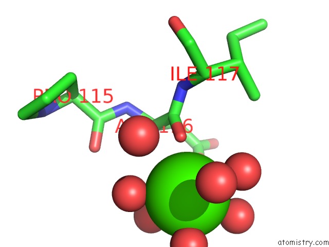
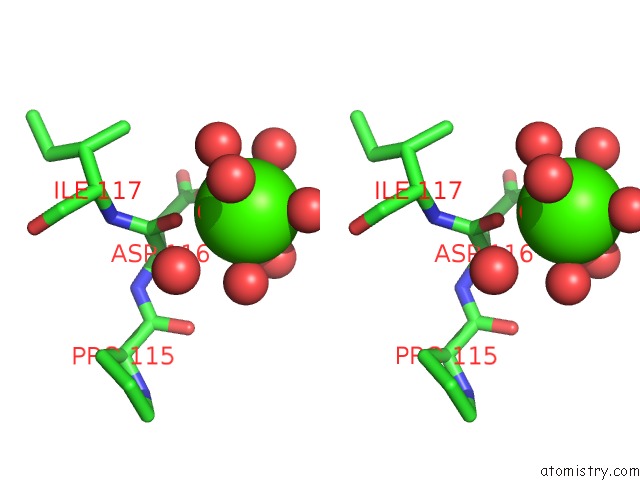
Calcium binding site 5 out of 6 in 2e85
Go back to
Calcium binding site 5 out
of 6 in the Crystal Structure of the Hydrogenase 3 Maturation Protease
Mono view
Stereo pair view
Mono view
Stereo pair view
A full contact list of Calcium with other atoms in the Ca binding
site number 5 of Crystal Structure of the Hydrogenase 3 Maturation Protease within 5.0Å range:
|
Calcium binding site 6 out of 6 in 2e85
Go back to
Calcium binding site 6 out
of 6 in the Crystal Structure of the Hydrogenase 3 Maturation Protease
Mono view
Stereo pair view
Mono view
Stereo pair view
A full contact list of Calcium with other atoms in the Ca binding
site number 6 of Crystal Structure of the Hydrogenase 3 Maturation Protease within 5.0Å range:
|
Reference:
T.Kumarevel,
T.Tanaka,
Y.Bessho,
A.Shinkai,
S.Yokoyama.
Crystal Structure of Hydrogenase Maturating Endopeptidase Hyci From Escherichia Coli Biochem.Biophys.Res.Commun. V. 389 310 2009.
ISSN: ISSN 0006-291X
PubMed: 19720045
DOI: 10.1016/J.BBRC.2009.08.135
Page generated: Fri Jul 12 10:07:54 2024
ISSN: ISSN 0006-291X
PubMed: 19720045
DOI: 10.1016/J.BBRC.2009.08.135
Last articles
Zn in 9MJ5Zn in 9HNW
Zn in 9G0L
Zn in 9FNE
Zn in 9DZN
Zn in 9E0I
Zn in 9D32
Zn in 9DAK
Zn in 8ZXC
Zn in 8ZUF