Calcium »
PDB 2eex-2f3z »
2ero »
Calcium in PDB 2ero: Crystal Structure of Vascular Apoptosis-Inducing Protein- 1(Orthorhombic Crystal Form)
Protein crystallography data
The structure of Crystal Structure of Vascular Apoptosis-Inducing Protein- 1(Orthorhombic Crystal Form), PDB code: 2ero
was solved by
S.Takeda,
T.Igarashi,
S.Araki,
with X-Ray Crystallography technique. A brief refinement statistics is given in the table below:
| Resolution Low / High (Å) | 50.00 / 2.50 |
| Space group | P 21 21 21 |
| Cell size a, b, c (Å), α, β, γ (°) | 86.710, 93.270, 137.740, 90.00, 90.00, 90.00 |
| R / Rfree (%) | 21.2 / 25.8 |
Other elements in 2ero:
The structure of Crystal Structure of Vascular Apoptosis-Inducing Protein- 1(Orthorhombic Crystal Form) also contains other interesting chemical elements:
| Cobalt | (Co) | 1 atom |
| Zinc | (Zn) | 2 atoms |
Calcium Binding Sites:
The binding sites of Calcium atom in the Crystal Structure of Vascular Apoptosis-Inducing Protein- 1(Orthorhombic Crystal Form)
(pdb code 2ero). This binding sites where shown within
5.0 Angstroms radius around Calcium atom.
In total 4 binding sites of Calcium where determined in the Crystal Structure of Vascular Apoptosis-Inducing Protein- 1(Orthorhombic Crystal Form), PDB code: 2ero:
Jump to Calcium binding site number: 1; 2; 3; 4;
In total 4 binding sites of Calcium where determined in the Crystal Structure of Vascular Apoptosis-Inducing Protein- 1(Orthorhombic Crystal Form), PDB code: 2ero:
Jump to Calcium binding site number: 1; 2; 3; 4;
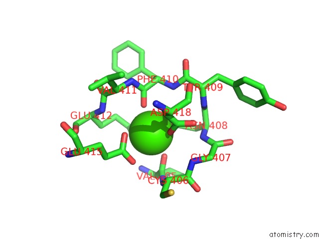
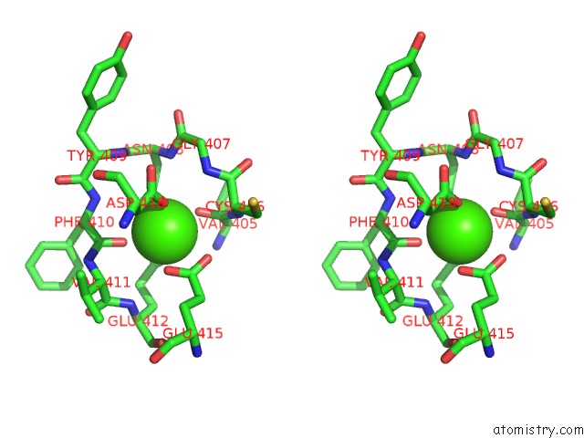
Calcium binding site 1 out of 4 in 2ero
Go back to
Calcium binding site 1 out
of 4 in the Crystal Structure of Vascular Apoptosis-Inducing Protein- 1(Orthorhombic Crystal Form)
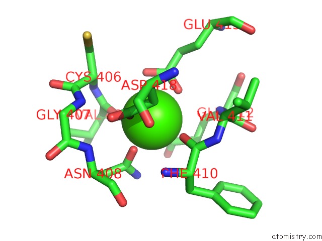
Mono view
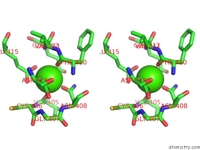
Stereo pair view
Mono view
Stereo pair view
A full contact list of Calcium with other atoms in the Ca binding
site number 1 of Crystal Structure of Vascular Apoptosis-Inducing Protein- 1(Orthorhombic Crystal Form) within 5.0Å range:
|
Calcium binding site 2 out of 4 in 2ero
Go back to
Calcium binding site 2 out
of 4 in the Crystal Structure of Vascular Apoptosis-Inducing Protein- 1(Orthorhombic Crystal Form)
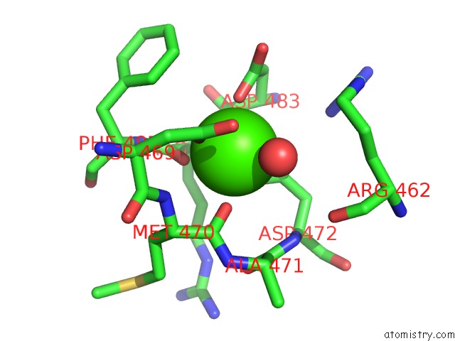
Mono view
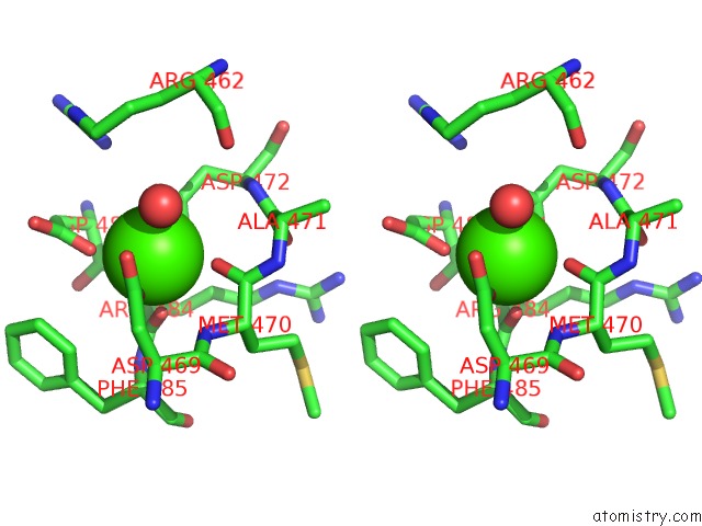
Stereo pair view
Mono view
Stereo pair view
A full contact list of Calcium with other atoms in the Ca binding
site number 2 of Crystal Structure of Vascular Apoptosis-Inducing Protein- 1(Orthorhombic Crystal Form) within 5.0Å range:
|
Calcium binding site 3 out of 4 in 2ero
Go back to
Calcium binding site 3 out
of 4 in the Crystal Structure of Vascular Apoptosis-Inducing Protein- 1(Orthorhombic Crystal Form)
Mono view
Stereo pair view
Mono view
Stereo pair view
A full contact list of Calcium with other atoms in the Ca binding
site number 3 of Crystal Structure of Vascular Apoptosis-Inducing Protein- 1(Orthorhombic Crystal Form) within 5.0Å range:
|
Calcium binding site 4 out of 4 in 2ero
Go back to
Calcium binding site 4 out
of 4 in the Crystal Structure of Vascular Apoptosis-Inducing Protein- 1(Orthorhombic Crystal Form)
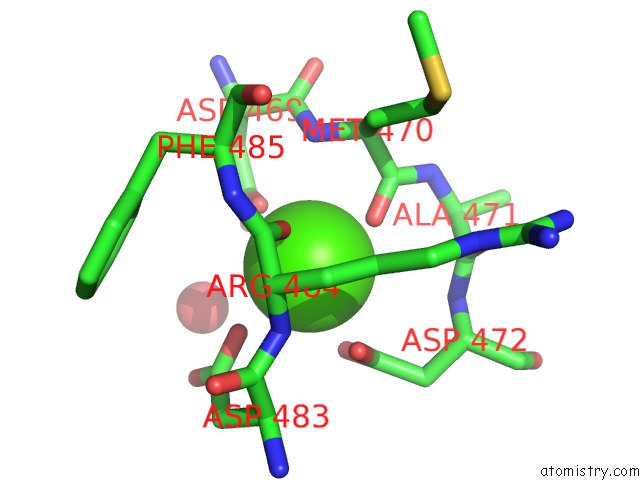
Mono view
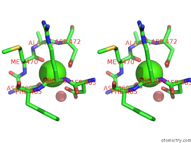
Stereo pair view
Mono view
Stereo pair view
A full contact list of Calcium with other atoms in the Ca binding
site number 4 of Crystal Structure of Vascular Apoptosis-Inducing Protein- 1(Orthorhombic Crystal Form) within 5.0Å range:
|
Reference:
S.Takeda,
T.Igarashi,
H.Mori,
S.Araki.
Crystal Structures of VAP1 Reveal Adams' Mdc Domain Architecture and Its Unique C-Shaped Scaffold Embo J. V. 25 2388 2006.
ISSN: ISSN 0261-4189
PubMed: 16688218
DOI: 10.1038/SJ.EMBOJ.7601131
Page generated: Tue Jul 8 05:22:42 2025
ISSN: ISSN 0261-4189
PubMed: 16688218
DOI: 10.1038/SJ.EMBOJ.7601131
Last articles
Ca in 7KB8Ca in 7KB6
Ca in 7KAD
Ca in 7K9T
Ca in 7KAA
Ca in 7K9Q
Ca in 7K9O
Ca in 7K9N
Ca in 7K6C
Ca in 7K96