Calcium »
PDB 2eex-2f3z »
2eu8 »
Calcium in PDB 2eu8: Crystal Structure of A Thermostable Mutant of Bacillus Subtilis Adenylate Kinase (Q199R)
Enzymatic activity of Crystal Structure of A Thermostable Mutant of Bacillus Subtilis Adenylate Kinase (Q199R)
All present enzymatic activity of Crystal Structure of A Thermostable Mutant of Bacillus Subtilis Adenylate Kinase (Q199R):
2.7.4.3;
2.7.4.3;
Protein crystallography data
The structure of Crystal Structure of A Thermostable Mutant of Bacillus Subtilis Adenylate Kinase (Q199R), PDB code: 2eu8
was solved by
S.Chen,
Y.Shamoo,
with X-Ray Crystallography technique. A brief refinement statistics is given in the table below:
| Resolution Low / High (Å) | 37.57 / 1.80 |
| Space group | P 1 21 1 |
| Cell size a, b, c (Å), α, β, γ (°) | 34.690, 75.140, 77.350, 90.00, 98.13, 90.00 |
| R / Rfree (%) | 18.7 / 22.4 |
Other elements in 2eu8:
The structure of Crystal Structure of A Thermostable Mutant of Bacillus Subtilis Adenylate Kinase (Q199R) also contains other interesting chemical elements:
| Magnesium | (Mg) | 2 atoms |
| Zinc | (Zn) | 2 atoms |
Calcium Binding Sites:
The binding sites of Calcium atom in the Crystal Structure of A Thermostable Mutant of Bacillus Subtilis Adenylate Kinase (Q199R)
(pdb code 2eu8). This binding sites where shown within
5.0 Angstroms radius around Calcium atom.
In total only one binding site of Calcium was determined in the Crystal Structure of A Thermostable Mutant of Bacillus Subtilis Adenylate Kinase (Q199R), PDB code: 2eu8:
In total only one binding site of Calcium was determined in the Crystal Structure of A Thermostable Mutant of Bacillus Subtilis Adenylate Kinase (Q199R), PDB code: 2eu8:
Calcium binding site 1 out of 1 in 2eu8
Go back to
Calcium binding site 1 out
of 1 in the Crystal Structure of A Thermostable Mutant of Bacillus Subtilis Adenylate Kinase (Q199R)
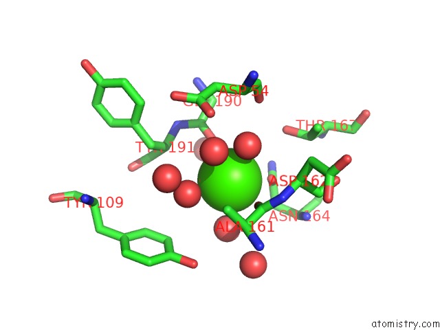
Mono view
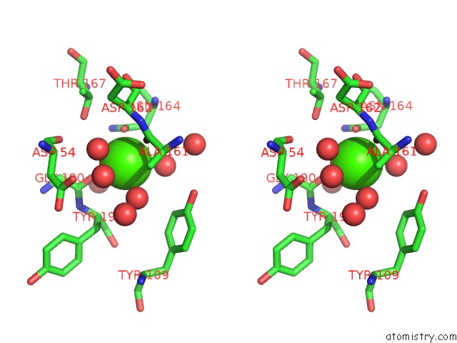
Stereo pair view
Mono view
Stereo pair view
A full contact list of Calcium with other atoms in the Ca binding
site number 1 of Crystal Structure of A Thermostable Mutant of Bacillus Subtilis Adenylate Kinase (Q199R) within 5.0Å range:
|
Reference:
R.Counago,
S.Chen,
Y.Shamoo.
In Vivo Molecular Evolution Reveals Biophysical Origins of Organismal Fitness. Mol.Cell V. 22 441 2006.
ISSN: ISSN 1097-2765
PubMed: 16713575
DOI: 10.1016/J.MOLCEL.2006.04.012
Page generated: Fri Jul 12 10:17:52 2024
ISSN: ISSN 1097-2765
PubMed: 16713575
DOI: 10.1016/J.MOLCEL.2006.04.012
Last articles
Zn in 9MJ5Zn in 9HNW
Zn in 9G0L
Zn in 9FNE
Zn in 9DZN
Zn in 9E0I
Zn in 9D32
Zn in 9DAK
Zn in 8ZXC
Zn in 8ZUF