Calcium »
PDB 2eex-2f3z »
2euf »
Calcium in PDB 2euf: X-Ray Structure of Human CDK6-Vcyclin in Complex with the Inhibitor PD0332991
Enzymatic activity of X-Ray Structure of Human CDK6-Vcyclin in Complex with the Inhibitor PD0332991
All present enzymatic activity of X-Ray Structure of Human CDK6-Vcyclin in Complex with the Inhibitor PD0332991:
2.7.1.37;
2.7.1.37;
Protein crystallography data
The structure of X-Ray Structure of Human CDK6-Vcyclin in Complex with the Inhibitor PD0332991, PDB code: 2euf
was solved by
U.Schulze-Gahmen,
H.Lu,
with X-Ray Crystallography technique. A brief refinement statistics is given in the table below:
| Resolution Low / High (Å) | 20.00 / 3.00 |
| Space group | P 65 2 2 |
| Cell size a, b, c (Å), α, β, γ (°) | 71.146, 71.146, 446.876, 90.00, 90.00, 120.00 |
| R / Rfree (%) | 22.9 / 30.6 |
Calcium Binding Sites:
The binding sites of Calcium atom in the X-Ray Structure of Human CDK6-Vcyclin in Complex with the Inhibitor PD0332991
(pdb code 2euf). This binding sites where shown within
5.0 Angstroms radius around Calcium atom.
In total only one binding site of Calcium was determined in the X-Ray Structure of Human CDK6-Vcyclin in Complex with the Inhibitor PD0332991, PDB code: 2euf:
In total only one binding site of Calcium was determined in the X-Ray Structure of Human CDK6-Vcyclin in Complex with the Inhibitor PD0332991, PDB code: 2euf:
Calcium binding site 1 out of 1 in 2euf
Go back to
Calcium binding site 1 out
of 1 in the X-Ray Structure of Human CDK6-Vcyclin in Complex with the Inhibitor PD0332991
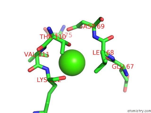
Mono view
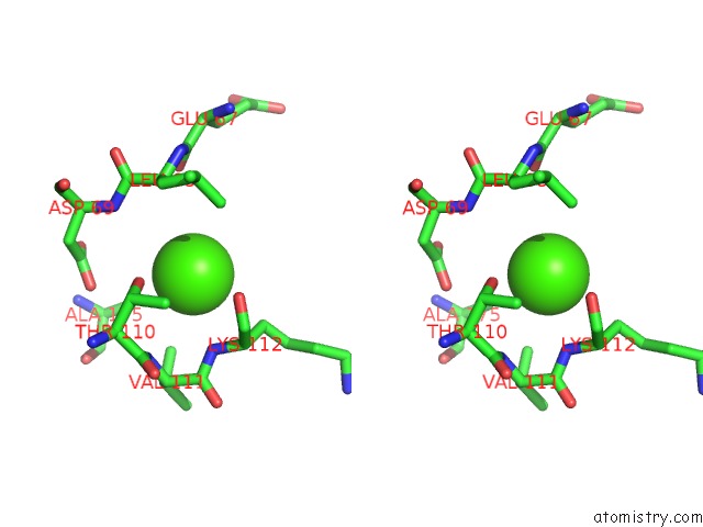
Stereo pair view
Mono view
Stereo pair view
A full contact list of Calcium with other atoms in the Ca binding
site number 1 of X-Ray Structure of Human CDK6-Vcyclin in Complex with the Inhibitor PD0332991 within 5.0Å range:
|
Reference:
H.Lu,
U.Schulze-Gahmen.
Toward Understanding the Structural Basis of Cyclin-Dependent Kinase 6 Specific Inhibition. J.Med.Chem. V. 49 3826 2006.
ISSN: ISSN 0022-2623
PubMed: 16789739
DOI: 10.1021/JM0600388
Page generated: Fri Jul 12 10:17:53 2024
ISSN: ISSN 0022-2623
PubMed: 16789739
DOI: 10.1021/JM0600388
Last articles
Zn in 9J0NZn in 9J0O
Zn in 9J0P
Zn in 9FJX
Zn in 9EKB
Zn in 9C0F
Zn in 9CAH
Zn in 9CH0
Zn in 9CH3
Zn in 9CH1