Calcium »
PDB 2eex-2f3z »
2exi »
Calcium in PDB 2exi: Structure of the FAMILY43 Beta-Xylosidase D15G Mutant From Geobacillus Stearothermophilus
Enzymatic activity of Structure of the FAMILY43 Beta-Xylosidase D15G Mutant From Geobacillus Stearothermophilus
All present enzymatic activity of Structure of the FAMILY43 Beta-Xylosidase D15G Mutant From Geobacillus Stearothermophilus:
3.2.1.37;
3.2.1.37;
Protein crystallography data
The structure of Structure of the FAMILY43 Beta-Xylosidase D15G Mutant From Geobacillus Stearothermophilus, PDB code: 2exi
was solved by
C.Brux,
K.Niefind,
D.Shallom-Shezifi,
Y.Shoham,
D.Schomburg,
with X-Ray Crystallography technique. A brief refinement statistics is given in the table below:
| Resolution Low / High (Å) | 20.00 / 2.15 |
| Space group | P 43 21 2 |
| Cell size a, b, c (Å), α, β, γ (°) | 140.175, 140.175, 232.322, 90.00, 90.00, 90.00 |
| R / Rfree (%) | 19.9 / 27.6 |
Calcium Binding Sites:
The binding sites of Calcium atom in the Structure of the FAMILY43 Beta-Xylosidase D15G Mutant From Geobacillus Stearothermophilus
(pdb code 2exi). This binding sites where shown within
5.0 Angstroms radius around Calcium atom.
In total 4 binding sites of Calcium where determined in the Structure of the FAMILY43 Beta-Xylosidase D15G Mutant From Geobacillus Stearothermophilus, PDB code: 2exi:
Jump to Calcium binding site number: 1; 2; 3; 4;
In total 4 binding sites of Calcium where determined in the Structure of the FAMILY43 Beta-Xylosidase D15G Mutant From Geobacillus Stearothermophilus, PDB code: 2exi:
Jump to Calcium binding site number: 1; 2; 3; 4;
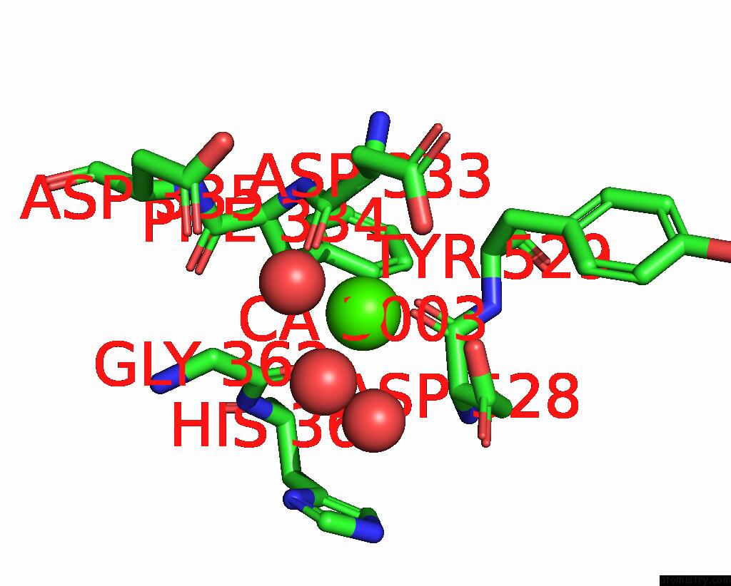
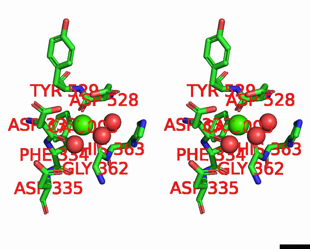
Calcium binding site 1 out of 4 in 2exi
Go back to
Calcium binding site 1 out
of 4 in the Structure of the FAMILY43 Beta-Xylosidase D15G Mutant From Geobacillus Stearothermophilus
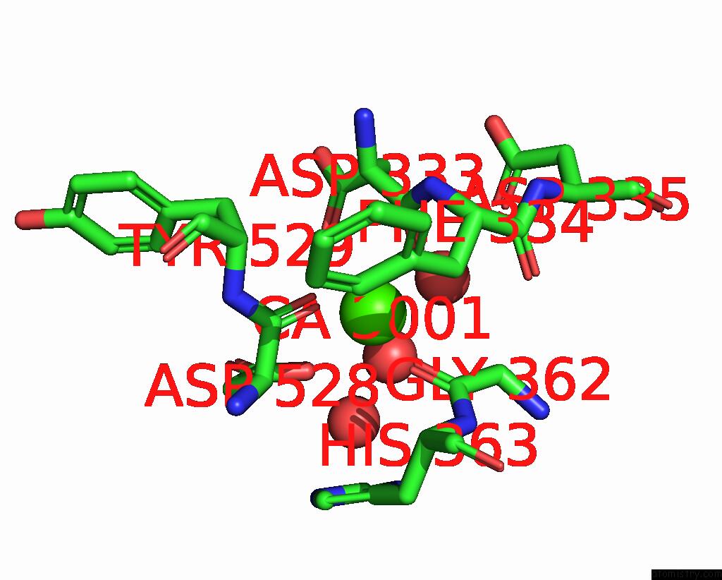
Mono view
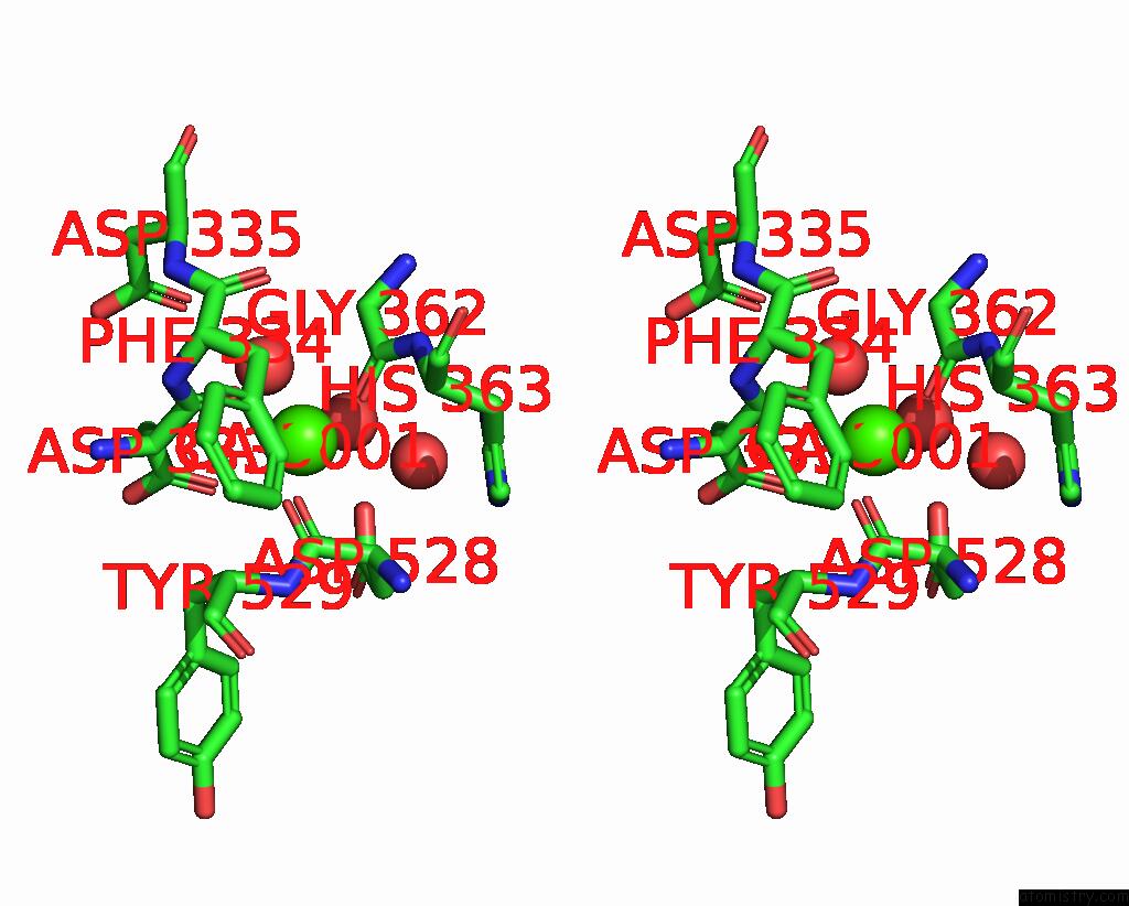
Stereo pair view
Mono view
Stereo pair view
A full contact list of Calcium with other atoms in the Ca binding
site number 1 of Structure of the FAMILY43 Beta-Xylosidase D15G Mutant From Geobacillus Stearothermophilus within 5.0Å range:
|
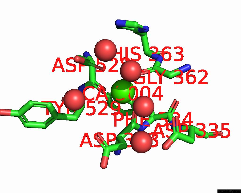
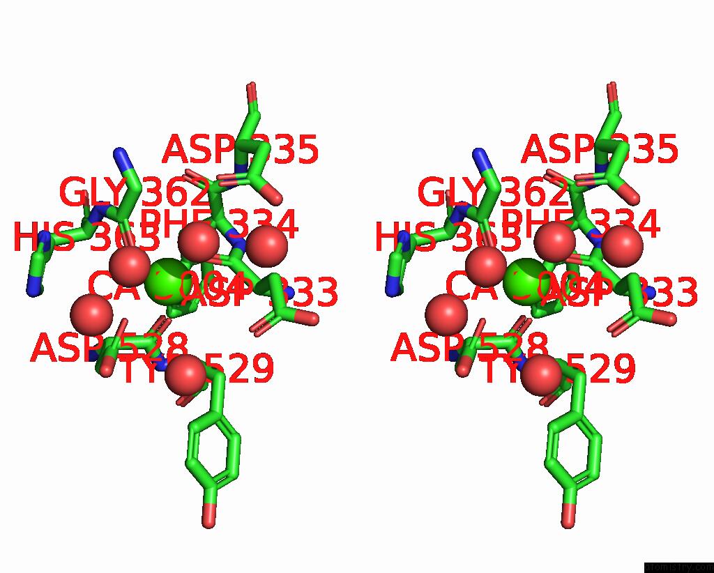
Calcium binding site 2 out of 4 in 2exi
Go back to
Calcium binding site 2 out
of 4 in the Structure of the FAMILY43 Beta-Xylosidase D15G Mutant From Geobacillus Stearothermophilus
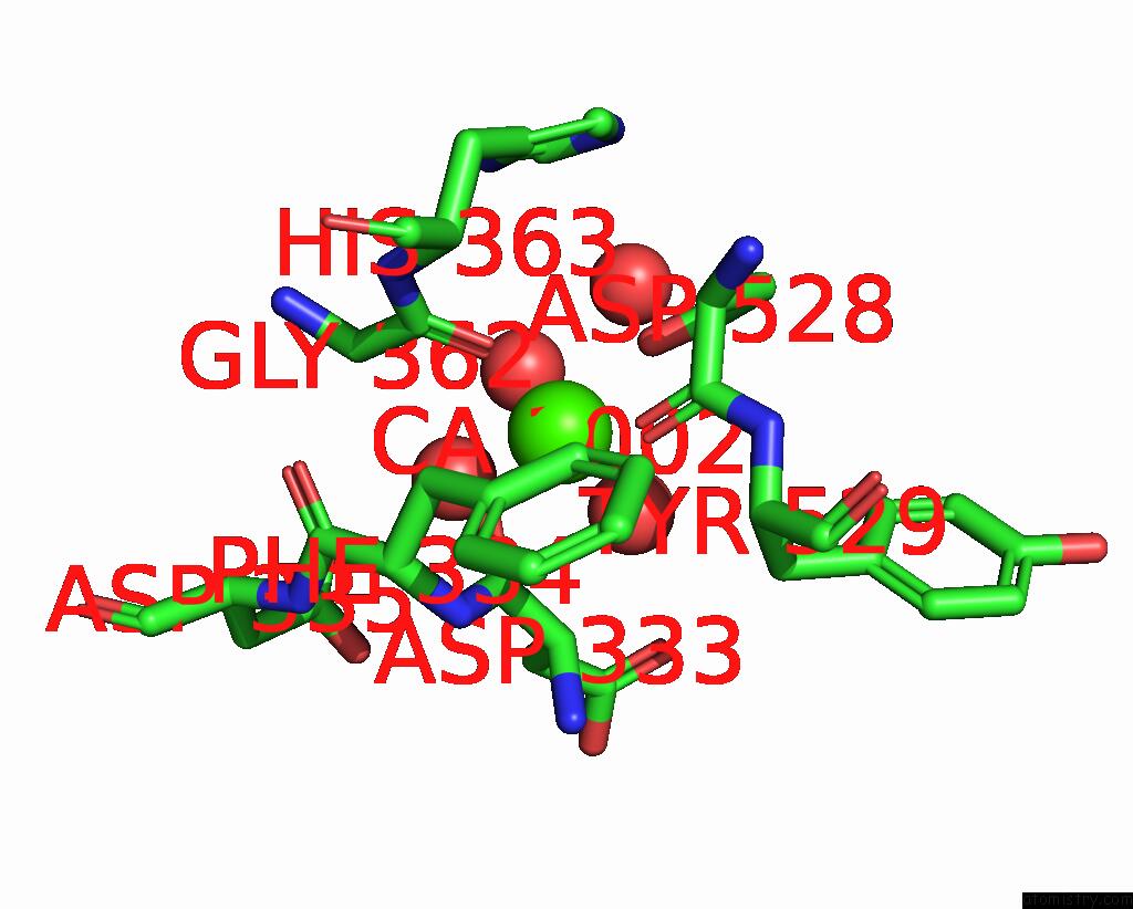
Mono view
Stereo pair view
Mono view
Stereo pair view
A full contact list of Calcium with other atoms in the Ca binding
site number 2 of Structure of the FAMILY43 Beta-Xylosidase D15G Mutant From Geobacillus Stearothermophilus within 5.0Å range:
|
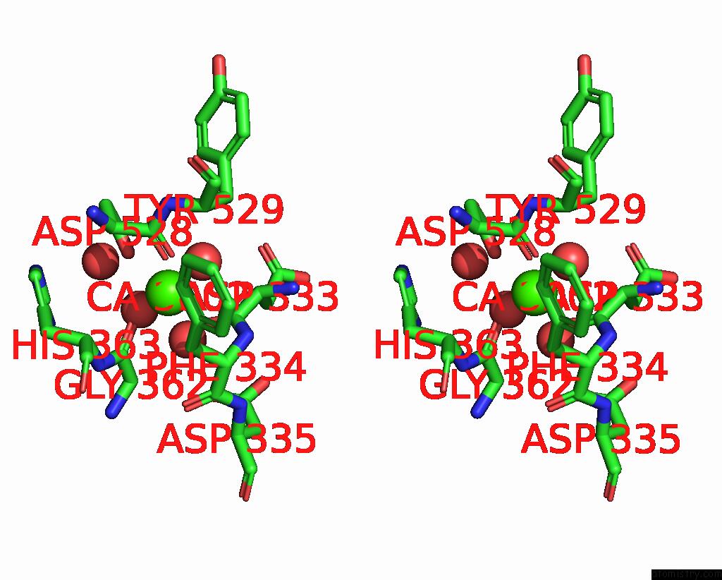
Calcium binding site 3 out of 4 in 2exi
Go back to
Calcium binding site 3 out
of 4 in the Structure of the FAMILY43 Beta-Xylosidase D15G Mutant From Geobacillus Stearothermophilus
Mono view
Stereo pair view
Mono view
Stereo pair view
A full contact list of Calcium with other atoms in the Ca binding
site number 3 of Structure of the FAMILY43 Beta-Xylosidase D15G Mutant From Geobacillus Stearothermophilus within 5.0Å range:
|
Calcium binding site 4 out of 4 in 2exi
Go back to
Calcium binding site 4 out
of 4 in the Structure of the FAMILY43 Beta-Xylosidase D15G Mutant From Geobacillus Stearothermophilus
Mono view
Stereo pair view
Mono view
Stereo pair view
A full contact list of Calcium with other atoms in the Ca binding
site number 4 of Structure of the FAMILY43 Beta-Xylosidase D15G Mutant From Geobacillus Stearothermophilus within 5.0Å range:
|
Reference:
C.Brux,
A.Ben-David,
D.Shallom-Shezifi,
M.Leon,
K.Niefind,
G.Shoham,
Y.Shoham,
D.Schomburg.
The Structure of An Inverting GH43 Beta-Xylosidase From Geobacillus Stearothermophilus with Its Substrate Reveals the Role of the Three Catalytic Residues. J.Mol.Biol. V. 359 97 2006.
ISSN: ISSN 0022-2836
PubMed: 16631196
DOI: 10.1016/J.JMB.2006.03.005
Page generated: Tue Jul 8 05:25:24 2025
ISSN: ISSN 0022-2836
PubMed: 16631196
DOI: 10.1016/J.JMB.2006.03.005
Last articles
Fe in 2YXOFe in 2YRS
Fe in 2YXC
Fe in 2YNM
Fe in 2YVJ
Fe in 2YP1
Fe in 2YU2
Fe in 2YU1
Fe in 2YQB
Fe in 2YOO